

Review
doi: 10.1016/S1054-3589(07)56016-3.
The viral etiology of AIDS-associated malignancies
Affiliations
- PMID: 18086422
- PMCID: PMC2149907
- DOI: 10.1016/S1054-3589(07)56016-3
Item in Clipboard
Review
The viral etiology of AIDS-associated malignancies
Adv Pharmacol.
2008.
No abstract available
Figures
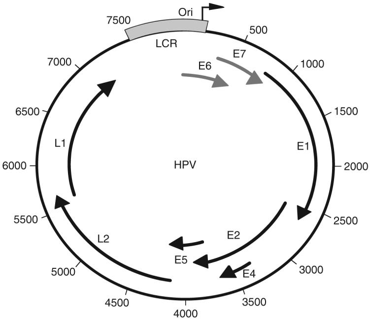
The HPV genome is a 7.9 kb double-stranded circular genome. The genome is controlled by a single keratin-dependent promoter element; the long control region (LCR; in blue). At the 3′ end of the LCR is the origin of replication (nucleotide position 1). E6, E7 (red), and E5 are viral oncogenes; E1 and E2 early genes encode replication proteins. The E4 ORF is actually expressed early and late in the viral life cycle. The late genes, L1 and L2, are the major and minor capsid genes, respectively. The viral protein functions are detailed in Table I.
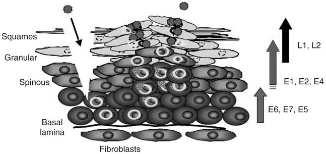
The HPV life cycle. Virions enter the stratified epithelium through a site of wounding, where they gain access to the mitotically active basal-layer keratinocytes. During the maintenance phase, expression of E6, E7, and E5 induces cell proliferation, and the viral genome is replicated extrachromosomally at low-copy number (5-50 copies per cell). As the cells differentiate, the expression level of E1, E2, and E4 increases in the spinous layer. A transition from theta to rolling-circle replication results in an increase in copy number up to 100-1000 copies per cell. Postamplification, high levels of L1 and L2 capsid genes are expressed and capsid assembly occurs in the granular and squamous layers of the stratified epithelium. Progeny virus is released by desquamation.
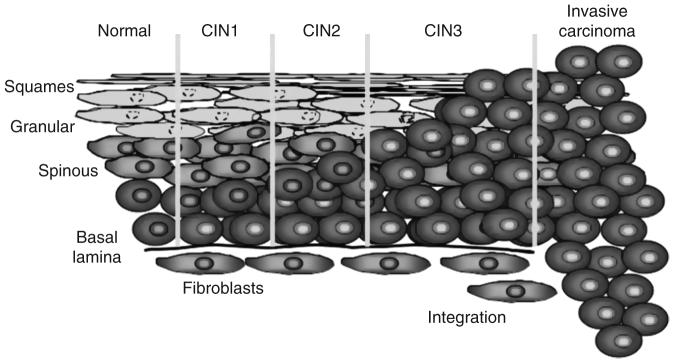
Progression from a benign cervical lesion to invasive cervical cancer. In the diagram, HPV-positive cells are depicted by yellow nuclei. Infection by oncogenic HPV types, especially HPV16, can cause formation of a benign wart, low or high-grade dysplasia- r. CIN 1 and CIN 2 designations are reversible forms of precancerous lesions and CIN 3 is irreversible. Carcinoma in situ occurs many years after an infection. This results from the effects of HPV genes, particularly those encoding E6 and E7, which are the two viral oncoproteins that are preferentially retained and expressed in cervical cancers by integration of the viral DNA into the host genome.
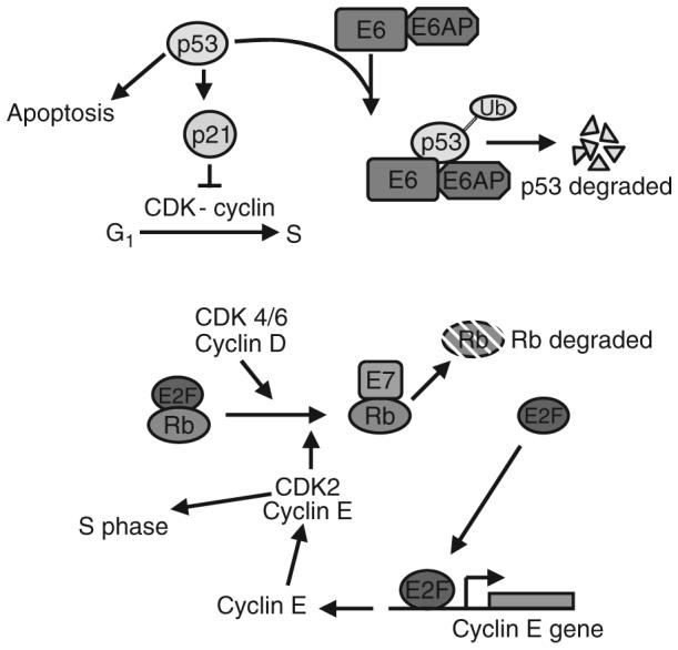
Diagram of the role of E6 and E7 in disregulation of the cell cycle. Expression of E6 leads to recruitment of E6AP (a ubiquitin ligase). This complex causes degradation of p53, which then inhibits the p21-dependent block of the G1 to S transition. Similarly, E7 binding to Rb displaces E2F, resulting in Rb’s degradation. E2F can then activate expression of cyclin E and other S-phase related gene products.
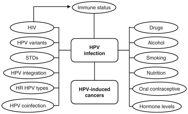
The rate of advancement of HPV lesions, from benign hyperplasia to carcinoma in situ, is affected by additional factors, which includes immunocompetence. HIV status, alcohol, drugs, smoking, oral contraceptives, and hormone levels influence HPV infection and progression of HPV-induced cancers. High-risk HPVs, HPV coinfection, variants, genome integration, and infection of other STDs affect the propensity for HPV-induced cancer to occur and progress.
Similar articles
-
Cancer in AIDS.Curr Opin Oncol. 2005 Sep;17(5):446. doi: 10.1097/01.cco.0000174042.37180.fb. Curr Opin Oncol. 2005. PMID: 16093793 No abstract available.
-
Kaposi's sarcoma findings.Science. 1995 Oct 6;270(5233):15. doi: 10.1126/science.270.5233.15b. Science. 1995. PMID: 7569942 No abstract available.
-
Liver involvement in AIDS-associated malignancies.J Hepatol. 1994 Dec;21(6):1145-6. doi: 10.1016/s0168-8278(05)80634-8. J Hepatol. 1994. PMID: 7699243 No abstract available.
-
Pathology of AIDS-related lymphomas and other AIDS-defining neoplasms.Eur J Cancer. 2001 Jul;37(10):1236-50. doi: 10.1016/s0959-8049(01)00103-4. Eur J Cancer. 2001. PMID: 11423256 Review. No abstract available.
-
AIDS-related malignancies.Nat Rev Cancer. 2002 May;2(5):373-82. doi: 10.1038/nrc797. Nat Rev Cancer. 2002. PMID: 12044013 Review.
Cited by
-
Human Immunodeficiency Virus-Associated Exosomes Promote Kaposi's Sarcoma-Associated Herpesvirus Infection via the Epidermal Growth Factor Receptor.J Virol. 2020 Apr 16;94(9):e01782-19. doi: 10.1128/JVI.01782-19. Print 2020 Apr 16. J Virol. 2020. PMID: 32051269 Free PMC article.
-
Clinical and pathological aspects of condyloma acuminatum - review of literature and case presentation.Rom J Morphol Embryol. 2021 Apr-Jun;62(2):369-383. doi: 10.47162/RJME.62.2.03. Rom J Morphol Embryol. 2021. PMID: 35024725 Free PMC article. Review.
-
Social and Structural Determinants of Cervical Health among Women Engaged in HIV Care.AIDS Behav. 2016 Sep;20(9):2101-9. doi: 10.1007/s10461-016-1345-6. AIDS Behav. 2016. PMID: 26955821 Free PMC article.
-
Epstein-Barr Virus, But Not Human Papillomavirus, Is Associated With Preinvasive and Invasive Ocular Surface Squamous Neoplasias in Zambian Patients.Front Oncol. 2022 Apr 14;12:864066. doi: 10.3389/fonc.2022.864066. eCollection 2022. Front Oncol. 2022. PMID: 35494029 Free PMC article.
-
Scrotal Kaposi's Sarcoma in HIV-negative patient: A case report and review of the literature.Turk J Urol. 2018 Mar;44(2):182-184. doi: 10.5152/tud.2017.68366. Epub 2017 Dec 11. Turk J Urol. 2018. PMID: 29511591 Free PMC article.
References
-
- Aaltonen LM, Cajanus S, Back L, Nieminen P, Paavonen J, Ranki A. Extralaryngeal HPV infections in male patients with adult-onset laryngeal papillomatosis. Eur. Arch. Otorhinolaryngol. 2005;262(9):708–712. - PubMed
-
- Ablashi D, Chatlynne L, Cooper H, Thomas D, Yadav M, Norhanom AW, Chandana AK, Churdboonchart V, Kulpradist SA, Patnaik M, Liegmann K, Masood R, et al. Seroprevalence of human herpesvirus-8 (HHV-8) in countries of Southeast Asia compared to the USA, the Caribbean and Africa. Br. J. Cancer. 1999;81(5):893–897. - PMC - PubMed
-
- Aboulafia DM, Gibbons R. Penile cancer and human papilloma virus (HPV) in a human immunodeficiency virus (HIV)-infected patient. Cancer Invest. 2001;19(3):266–272. - PubMed
-
- ACS . Cancer facts and figures. American Cancer Society; 2006. www.cancer.org.
-
- Agha S, Kusanthan T. Equity in access to condoms in urban Zambia. Health Policy Plan. 2003;18(3):299–305. - PubMed
Publication types
MeSH terms
Grants and funding
LinkOut - more resources
Full Text Sources
Medical
