Comparative Study
. 2008 Mar;29(3):422-37.
doi: 10.1016/j.nbd.2007.10.005. Epub 2007 Nov 4.

Cholesterol retention in Alzheimer's brain is responsible for high beta- and gamma-secretase activities and Abeta production

Affiliations
Comparative Study

Cholesterol retention in Alzheimer's brain is responsible for high beta- and gamma-secretase activities and Abeta production

Huaqi Xiong et al. Neurobiol Dis. 2008 Mar.

Abstract

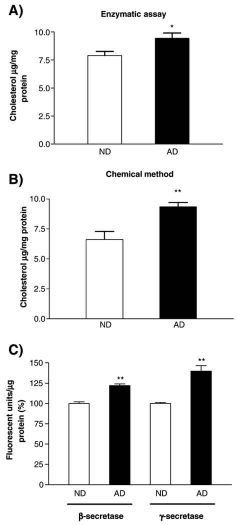
Alzheimer's disease (AD) is characterized by overproduction of A beta derived from APP cleavage via beta- and gamma-secretase pathway. Recent evidence has linked altered cholesterol metabolism to AD pathogenesis. In this study, we show that AD brain had significant cholesterol retention and high beta- and gamma-secretase activities as compared to age-matched non-demented controls (ND). Over one-half of AD patients had an apoE4 allele but none of the ND. beta- and gamma-secretase activities were significantly stimulated in vitro by 40 and 80 microM cholesterol in AD and ND brains, respectively. Both secretase activities in AD brain were more sensitive to cholesterol (40 microM) than those of ND (80 microM). Filipin-stained cholesterol overlapped with BACE and A beta in AD brain sections. Cholesterol (10-80 microM) added to N2a cultures significantly increased cellular cholesterol, beta- and gamma-secretase activities and A beta secretion. Similarly, addition of cholesterol (20-80 microM) to cell lysates stimulated both in vitro secretase activities. Ergosterol slightly decreased beta-secretase activity at 20-80 microM, but strongly inhibited gamma-secretase activity at 40 microM. Cholesterol depletion reduced cellular cholesterol, beta-secretase activity and A beta secretion. Transcription factor profiling shows that several key nuclear receptors involving cholesterol metabolism were significantly altered in AD brain, including decreased LXR-beta, PPAR and TR, and increased RXR. Treatment of N2a cells with LXR, RXR or PPAR agonists strongly stimulated cellular cholesterol efflux to HDL and reduced cellular cholesterol and beta-/gamma-secretase activities. This study provides direct evidence that cholesterol homeostasis is impaired in AD brain and suggests that altered levels or activities of nuclear receptors may contribute to cholesterol retention which likely enhances beta- and gamma-secretase activities and A beta production in human brain.


Figures

Fig. 1
Fig. 1
Detection of Aβ peptides and cholesterol in human brain samples. Brain sections were reacted with a primary 6E10 antibody and then with a secondary antibody conjugated with Alexa 568. Beta-amyloid is visualized as red color. Filipin is used to stain cholesterol in brain sections. Filipin-stained cholesterol is visualized as blue color. Overlapping staining of cholesterol and beta-amyloid shows blue-reddish haze color. Panels A and B are ND samples. Panels C–F are AD brain samples. All of the images were taken at 200× magnifications.
Fig. 2
Fig. 2
Levels of cholesterol and the activities of β- and γ-secretases in ND and AD brain samples. Panel A: Enzymatic assay shows that the level of cholesterol in AD is significantly higher (19.37%↑) than that in ND brain samples (t test, * p=0.0104). Panel B shows the results of chemical method analysis of total brain cholesterol. AD brains have significantly higher cholesterol (41.3%↑) than ND controls (t test, p<0.01) Panel C: Brain tissue lysates were prepared for β- and γ-secretase assays following manufacturer’s instructions as described in Materials and methods. The activities of both β-secretase and γ-secretases are significantly higher in AD than in ND brain samples (t test, ** p<0.01).
Fig. 3
Fig. 3
Effects of exogenous cholesterol on β- and γ-secretase activities in human brain tissue lysates. Panel A shows that 80 µM cholesterol (Cho) strongly stimulated β-secretase activity in ND brain lysates (One-way ANOVA, ***p<0.0001); while 40 µM and 80 µM cholesterol significantly enhanced β-secretase activity in AD brain lysates (One-way ANOVA, ***p<0.0001), respectively. Panel B shows that 80 µM cholesterol strongly stimulated γ-secretase activity in ND samples (One-way ANOVA, ***p<0.0001); while 40 µM cholesterol significantly stimulated γ-secretase activity in AD samples (One-way ANOVA, ***p<0.0001). Cholesterol at 20 µM concentration did not affect β-secretase activity, but inhibited γ-secretase activity in AD brain samples (***p<0.0001).
Fig. 4
Fig. 4
Immunostaining for BACE, beta-amyloid, and cholesterol. AD brain sections were immuno-reacted with a primary anti-human BACE antibody and then with a secondary antibody conjugated with Alexa 568. BACE is visualized as red color. Panels A and C are BACE staining for AD brain sections. Panels B and D are sister slides of panels A and C sections (respectively) and were reacted with a primary beta-amyloid antibody 6E10 and then with a secondary antibody conjugated with Alexa 568. The beta-amyloid in Panels B and D is also visualized as red color. Panels E and F show double-staining of AD brains with filipin for cholesterol (blue color) and BACE (red color). Panels G and H show BACE staining for N2a and K269 cells. All of the images were taken at 200× magnifications.
Fig. 5
Fig. 5
Effects of exogenous cholesterol in culture media on β- and γ-secretase activities in N2a cells. Different concentrations of exogenous cholesterol (Cho) were added to the culture media of N2a cells and incubated for 1, 2, 4, and 6 h. Cells were harvested for lysate preparations. Panel A: Cellular cholesterol was significantly higher in cholesterol-treated cells than in controls (One-way ANOVA, *p<0.05). Panels B and C: The in vitro assays show that cholesterol added to culture media significantly stimulated β- and γ-secretase activities in N2a cells (One-way ANOVA, *p<0.05). Panel D: Aβ1–40 peptides released into the media from cholesterol-treated cells were significantly increased at all the time points compared to controls (One-way ANOVA, ***p<0.0005, ***p<0.0001).
Fig. 6
Fig. 6
In vitro β- and γ-secretase activity assays in the presence of exogenous cholesterol. Cell lysates were prepared from N2a cells following manufacturer’s instructions as described in Materials and methods. Different concentrations of exogenous cholesterol or ergosterol were added to the in vitro assays for β- or γ-secretase activities, respectively. Panel A: The activity of β-secretase was significantly stimulated in the presence of 20–80 µM cholesterol as compared to controls (One-way ANOVA, *p=0.0198). Ergosterol at 20, 40 and 80 µM slightly decreased β-secretase activity. Panel B: Cholesterol at 20, 40 and 80 µM strongly stimulated γ-secretase activity (One-way ANOVA, ***p<0.0001). It appears that 20–40 µM cholesterol was stronger in stimulating γ-secretase activity than 80 µM cholesterol. Ergosterol at 40 µM strongly decreased γ-secretase activity (One-way ANOVA, ***p<0.0001). Panel C: The activities of β- and γ-secretases were significantly inhibited by p-secretase inhibitor III and γ-secretase inhibitor IV, respectively (One-way ANOVA, ***p<0.0001). The inhibitors inhibited 25–30% of β- and γ-secretase activities no matter in the presence or absence of cholesterol.
Fig. 7
Fig. 7
Effects of cholesterol depletion on levels of cellular cholesterol, β-secretase activity and Aβ production in N2a cells. Panel A shows that cellular cholesterol was significantly decreased at 4 h post-treatment with 2.5 mM cyclodextran (CDT) (One-way ANOVA, * p=0.0442), returned to control level at 8, 12, and 24 h. Panel B shows that β-secretase activity in CDT-treated cells was reduced at 4 h (One-way ANOVA, *p=0.0215), but returned to control levels at 8, 12 and 24 h. Aβ1–40 peptides released from CDT-treated cells were significantly lower at 4 h (One-way ANOVA, **p<0.0009), but slightly higher at 8, 12 and 24 h than controls (Panel C).
Fig. 8
Fig. 8
Activation of transcription factors in ND vs. AD brain samples. Panels A and B show transcription factor (TF) profiling using TranSignal protein/ DNA array blot I analysis (Panomics Inc.). The boxed transcription factors are involved in the regulation of lipid and cholesterol metabolism in cells. The binding activities of PPAR, TR, and VDR were decreased over 2-fold in AD; while the activities of RXR, ARE, ERE, and PRE were increased over 2-fold in AD brain samples as compared to ND brain samples (t test, p<0.05). The levels of sterol regulatory binding element transcription factor (SREBF or SREBP), GR and RAR were similar in ND and AD samples. Panel C shows the fold changes for PPAR, RXR, and TR in AD brain samples. The activity of the relevant transcription factor in ND brain samples was set as 1.
Fig. 9
Fig. 9
Level of LXR-β in ND vs. AD brain samples. Semi-quantitative RT-PCR was carried out to analyze the expression of LXR-β in ND and AD brain samples. The levels of LXR-β were generally lower in AD than in ND brain samples. The graph shows quantitative densitometry analysis of LXR-β expression in ND vs. AD brain samples normalized against an internal control β-actin. The average level of LXR-β in AD is 1.56-fold higher than that in ND (t test, **p<0.01).
Fig. 10
Fig. 10
Effects of LXR and RXR agonists on cholesterol efflux and β- and γ-secretase activities in cells. Cholesterol assays using K269 cells (Panel A) show that more cholesterol (41%) was transported to HDL after 8 h incubation of the cells with 5 µM LXR agonist TO901317 (TO) (One-way ANOVA, ***p< 0.0001). In the absence of TO, only a small fraction of cholesterol (22%) was shed to culture media compared to controls (One-way ANOVA, ***p< 0.0001). Panel B shows that cholesterol levels were decreased in N2a cells treated with LXR or/and RXR compared to controls (One-way ANOVA, *p<0.05). Since cellular cholesterol was decreased in LXR- and RXR-treated N2a cells, β- and γ-secretase activities were significantly reduced in the cells (One-way ANOVA, *p<0.05, **p<0.01) (Panels C and D).
Fig. 11
Fig. 11
Effects of PPAR-γ agonist troglitazone on β- and γ-secretase activities in N2a cells. Both β-secretase (Panel A) and γ-secretase (Panel B) activities were significantly decreased in N2a cells treated with 10 µM troglitazone (TROG) as compared to controls (One-way ANOVA, Panel A, *p=0.0108; Panel B, ***p<0.0001) at 24 and 36h post-treatment

Similar articles

Cited by

References

    1. Abad-Rodriguez J, Ledesma MD, Craessaerts K, Perga S, Medina M, Delacourte A, Dingwall C, De Strooper B, Dotti CG. Neuronal membrane cholesterol loss enhances amyloid peptide generation. J. Cell Biol. 2004;167(5):953–960. - PMC - PubMed
    1. Abulrob A, Tauskela JS, Mealing G, Brunette E, Faid K, Stanimirovic D. Protection by cholesterol-extracting cyclodextrins: a role for N-methyl-D-aspartate receptor redistribution. J. Neurochem. 2005;92(6):1477–1486. - PubMed
    1. Akiyama TE, Sakai S, Lambert G, Nicol CJ, Matsusue K, Pimprale S, Lee YH, Ricote M, Glass CK, Brewer HB, Jr, Gonzalez FJ. Conditional disruption of the peroxisome proliferator-activated receptor gamma gene in mice results in lowered expression of ABCA1, ABCG1, and apoE in macrophages and reduced cholesterol efflux. Mol. Cell. Biol. 2002;22(8):2607–2619. - PMC - PubMed
    1. Bjorkhem I, Meaney S. Brain cholesterol: long secret life behind a barrier. Arterioscler. Thromb. Vasc. Biol. 2004;24:806–815. - PubMed
    1. Cordy JM, Hooper NM, Turner AJ. Exclusively targeting beta-secretase to lipid rafts by GPI-anchor addition up-regulates beta-site processing of the amyloid precursor protein. Proc. Natl. Acad. Sci. U. S. A. 2003;100(20):11735–11740. - PMC - PubMed

Publication types

Substances