Review
. 2008 Jan-Feb;18(1):11-21.
doi: 10.1684/ejd.2008.0304. Epub 2007 Dec 18.

Antimicrobial peptides in human skin disease

Affiliations
Review

Antimicrobial peptides in human skin disease

Kenshi Yamasaki et al. Eur J Dermatol. 2008 Jan-Feb.

Abstract

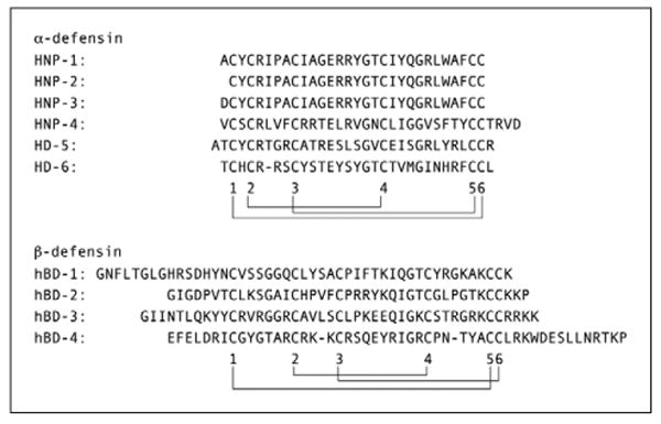
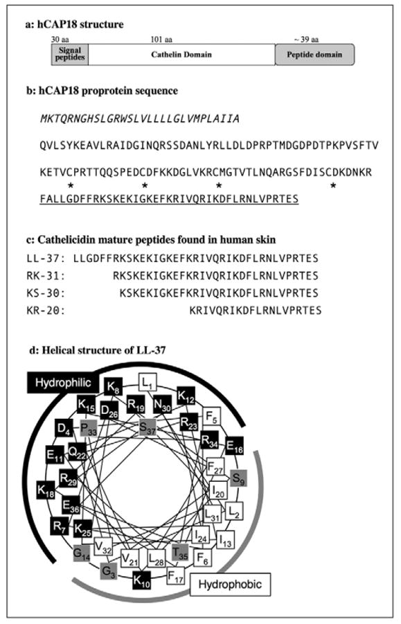
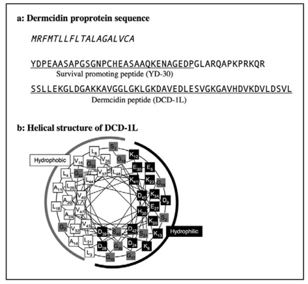
The skin continuously encounters microbial pathogens. To defend against this, cells of the epidermis and dermis have evolved several innate strategies to prevent infection. Antimicrobial peptides are one of the primary mechanisms used by the skin in the early stages of immune defense. In general, antimicrobial peptides have broad antibacterial activity against gram-positive and negative bacteria and also show antifungal and antiviral activity. The antimicrobial activity of most peptides occurs as a result of unique structural characteristics that enable them to disrupt the microbial membrane while leaving human cell membranes intact. However, antimicrobial peptides also act on host cells to stimulate cytokine production, cell migration, proliferation, maturation, and extracellular matrix synthesis. The production by human skin of antimicrobial peptides such as defensins and cathelicidins occurs constitutively but also greatly increases after infection, inflammation or injury. Some skin diseases show altered expression of antimicrobial peptides, partially explaining the pathophysiology of these diseases. Thus, current research suggests that understanding how antimicrobial peptides modify susceptibility to microbes, influence skin inflammation, and modify wound healing, provides greater insight into the pathophysiology of skin disorders and offers new therapeutic opportunities.


Figures

Figure 1
Figure 1. Human defensin peptide sequences
Alignment of the six human α-defensin peptides and four β-defensin peptides. The location of the six cysteins and the disulfide bond connectivity are indicated with digits and lines, respectively.
Figure 2
Figure 2. Human cathelicidin hCAP18 proprotein structure and sequence
A) Schematic representation of the hCAP18 proprotein structure. B) Italic characters indicate signal peptide, and mature peptide domain of LL-37 is underlined. Asterisks indicate four cysteins, which are conserved in the cathelicidin proprotein between species and form disulfide bonds in the cathelin domain. Postsecretory processing generates cathelicidin mature peptides although the sizes and sequences of mature peptides are varied and depend on tissues or cell-specific proteases. C) Four representative human skin cathelicidin peptides LL-37, RK-31, KS-30, and KR-20 are listed. d: α-helical wheel of LL-37. Black boxes indicate hydrophilic amino acids, white boxes indicate hydrophobic amino acids, and gray boxes are amphiphilic amino acids [165]. Numeric numbers indicate the order in the peptide. A black line indicates the hydrophilic side and a gray line indicates the hydrophobic side of the peptides.
Figure 3
Figure 3. Human Dermcidin amino acid sequence
A) Italic characters indicate the signal peptide, and functional peptide domains are underlined. Dermcidin proprotein generates at least two different peptides; surviving promoting peptide (YD-30) and dermcidin antimicrobial peptide (DCD-1L). B) -helical wheel of DCD-1L. Black boxes indicate hydrophilic amino acids, white boxes indicate hydrophobic amino acids, and gray boxes are amphiphilic amino acids [165]. Numeric numbers indicate the order in the peptide. A black line indicates the hydrophilic side and a gray line indicates the hydrophobic side of the peptides.

Similar articles

Cited by

References

    1. Gallo RL, Ono M, Povsic T, Page C, Eriksson E, Klagsbrun M, Bernfield M. Syndecans, cell surface heparan sulfate proteoglycans, are induced by a prolinerich antimicrobial peptide from wounds. Proc Natl Acad Sci USA. 1994;91:11035–9. - PMC - PubMed
    1. Peschel A, Otto M, Jack RW, Kalbacher H, Jung G, Gotz F. Inactivation of the dlt operon in Staphylococcus aureus confers sensitivity to defensins, protegrins, and other antimicrobial peptides. J Biol Chem. 1999;274:8405–10. - PubMed
    1. Kristian SA, Datta V, Weidenmaier C, Kansal R, Fedtke I, Peschel A, Gallo RL, Nizet V. D-alanylation of teichoic acids promotes group a streptococcus antimicrobial peptide resistance, neutrophil survival, and epithelial cell invasion. J Bacteriol. 2005;187:6719–25. - PMC - PubMed
    1. Larrick JW, Hirata M, Balint RF, Lee J, Zhong J, Wright SC. Human CAP18: a novel antimicrobial lipopolysaccharide-binding protein. Infect Immun. 1995;63:1291–7. - PMC - PubMed
    1. Ganz T, Weiss J. Antimicrobial peptides of phagocytes and epithelia. Semin Hematol. 1997;34:343–54. - PubMed

Publication types

MeSH terms

LinkOut - more resources