. 2008 Feb;36(3):938-49.
doi: 10.1093/nar/gkm1090. Epub 2007 Dec 17.

Tetrameric restriction enzymes: expansion to the GIY-YIG nuclease family

Affiliations

Tetrameric restriction enzymes: expansion to the GIY-YIG nuclease family

Giedrius Gasiunas et al. Nucleic Acids Res. 2008 Feb.

Abstract

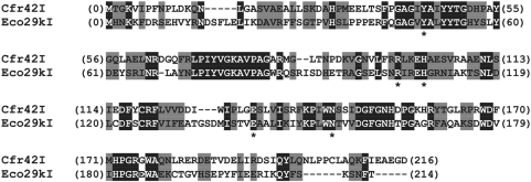
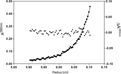
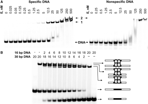
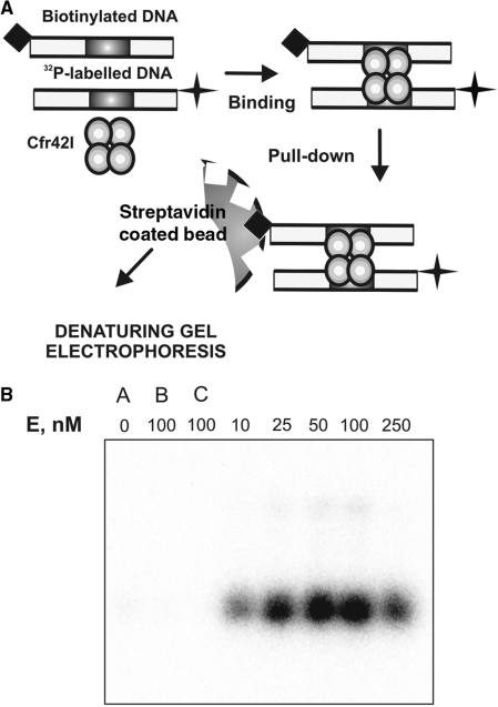
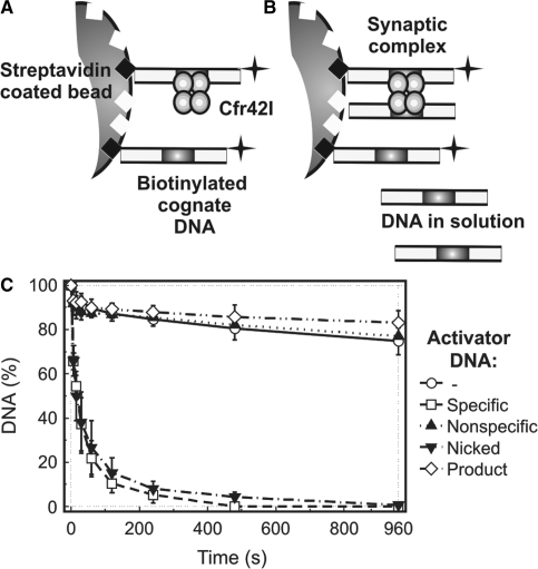
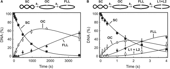
The GIY-YIG nuclease domain was originally identified in homing endonucleases and enzymes involved in DNA repair and recombination. Many of the GIY-YIG family enzymes are functional as monomers. We show here that the Cfr42I restriction endonuclease which belongs to the GIY-YIG family and recognizes the symmetric sequence 5'-CCGC/GG-3' ('/' indicates the cleavage site) is a tetramer in solution. Moreover, biochemical and kinetic studies provided here demonstrate that the Cfr42I tetramer is catalytically active only upon simultaneous binding of two copies of its recognition sequence. In that respect Cfr42I resembles the homotetrameric Type IIF restriction enzymes that belong to the distinct PD-(E/D)XK nuclease superfamily. Unlike the PD-(E/D)XK enzymes, the GIY-YIG nuclease Cfr42I accommodates an extremely wide selection of metal-ion cofactors, including Mg2+, Mn2+, Co2+, Zn2+, Ni2+, Cu2+ and Ca2+. To our knowledge, Cfr42I is the first tetrameric GIY-YIG family enzyme. Similar structural arrangement and phenotypes displayed by restriction enzymes of the PD-(E/D)XK and GIY-YIG nuclease families point to the functional significance of tetramerization.


Figures

Figure 1.
Figure 1.
Alignment of Cfr42I and Eco29kI sequences. Identical amino acid residues (32%) are in black boxes, similar residues (32%) are shaded grey. Asterisks (*) mark amino acid residues Y49, R104, H108, E142 and N154 of Eco29kI that are critical for its catalytic activity and presumably form the active site of Eco29kI (5).
Figure 2.
Figure 2.
Analytical ultracentrifugation of Cfr42I. Sedimentation diffusion equilibrium analysis of 3.2 µM Cfr42I (monomer) was performed at 18 000 rpm and 20°C in 0.01 M Tris-HCl pH 7.4, 0.2 M KCl, 0.1 mM EDTA. Filled squares denote the measured absorption (A280nm, left ordinate). The solid line shows the best least squares fit to the data for a single species with a molar mass of 99 ± 3 kg/mol (cf. (10)). Filled triangles denote the residues of that fit (ΔA280nm, right ordinate).
Figure 3.
Figure 3.
DNA binding by Cfr42I endonuclease. (A) DNA-binding analysis by gel mobility-shift assay. The reactions contained 1 nM of the 33P-labelled specific oligoduplex 30/30 or the non-specific oligonucleotide NS (see Table 1 for sequence details), and the protein at concentrations (in terms of nM tetramer) as indicated above each lane. After 15 min at room temperature, the samples were subjected to PAGE for 2 h and analysed as described in ‘Materials and Methods’. (B) Analysis of the Cfr42I-DNA complex. Cfr42I (12.5 nM of tetramer) was added to the 30/30 or 16/16 33P-labelled specific DNA duplexes (Table 1) alone or to the duplex mixtures in ratios varying from 9:1 to 1:9 keeping the total DNA concentration fixed at 20 nM. Concentrations of both duplexes in the reactions are indicated above each line. The extreme left and right gel lanes contained no protein. After 15 min at room temperature, the samples were subjected to PAGE for 3 h and analysed as described in ‘Materials and Methods’. The cartoons illustrate protein-DNA complexes that correspond to each band.
Figure 4.
Figure 4.
DNA synapsis by Cfr42I. (A) Schematic representation of the biotin pull-down assay. The reactions contained equimolar amounts of two specific 30 bp duplexes (10 nM each) with various amounts of Cfr42I. The first of the duplexes carried a biotin tag and the other was radiolabelled with 32P. After 5 min preincubation of enzyme and DNA, streptavidin-coated magnetic beads were added that adsorbed the biotin-tagged DNA. The beads were harvested and the amount of radiolabelled DNA associated with the beads was measured after denaturing PAGE. The radiolabelled DNA is pulled down with the beads only if Cfr42I forms complexes with two DNA molecules. (B) Results of the pull-down assay. Cfr42I concentrations were as indicated above each lane. The gel lanes marked ‘A’, ‘B’ and ‘C’ are control pull-down experiments performed in the absence of protein (lane ‘A’), in the presence of non-specific biotinylated DNA (lane ‘B’) or in the absence of biotinylated DNA (lane ‘C’).
Figure 5.
Figure 5.
Immobilized DNA cleavage by Cfr42I. Cartoons in (A) and (B) depict the experimental strategy (36). (A) Oligonucleotide duplex bio-30-30 (Table 1), which carries a biotin at the 5′-end and a 32P label at the 3′ end, is immobilized on the streptavidin-coated magnetic beads at low density. These reaction conditions prevent formation of synaptic complexes between tetrameric REase and two DNA molecules and thus reveal catalytic activity of enzyme bound to a single DNA site. (B) Alternatively, addition of non-biotinylated oligonucleotide duplex (activator DNA) into the reaction mixture enables formation of synaptic complexes between the enzyme, the immobilized DNA and the activator DNA in solution. (C) Experimental results. Upon addition of 25 nM of tetrameric Cfr42I, cleavage of the immobilized hairpin duplex (∼1 nM) was monitored by removing samples at timed intervals (open circles) and analysing them as described in ‘Materials and Methods’. Cleavage of immobilized substrate was also performed in the presence of various non-biotinylated activator DNAs: 50 nM non-specific duplex NS (filled triangles), 50 nM cognate duplex 30/30 (open squares), 50 nM product DNA 16/14p (open diamonds) and 50 nM cognate nicked duplex 30/(14p_16) (inverted filled triangles) (see Table 1 for oligonucleotide sequences). All data points are presented as mean values from ≥3 independent experiments ±1SD.
Figure 6.
Figure 6.
Cfr42I reactions on plasmids with one or two recognition sites. The reactions were performed by mixing solution of MgCl2 (final concentration 10 mM) with the preincubated mixture of enzyme and plasmid DNA (final concentrations 50 nM enzyme tetramer and 2.5 nM DNA). The plasmids were pBRCFR-1 (one Cfr42I site) for (A), and pBRCFR-2 (two Cfr42I sites) for (B). Samples were quenched with 6 M guanidinium chloride and analysed as described in ‘Materials and Methods’ to determine the amounts of the following forms of the DNA: supercoiled DNA (SC), filled squares; open-circular DNA (OC), open triangles; linear DNA cut at one Cfr42I site (FLL), filled triangles; and, only in (B), linear DNA cut at both Cfr42I sites (L1 + L2), open circles. Cartoons above the graphs depict the two-step and three-step consecutive reaction schemes that were used to quantify the one-site and two-site plasmid DNA cleavage data as described (19). The solid lines in panel (A) show the best least squares fit of the two-step reaction scheme to the one-site plasmid data that gave k1 = 0.0013 ± 0.0001 s−1 and k2 = 0.0004 ± 0.0001 s−1. The solid lines in panel (B) show the best fit of the three-step scheme to the two-site plasmid data that gave k1 = 0.56 ± 0.02 s−1, k2 = 0.56 ± 0.03 s−1 and k3 = 0.17 ± 0.02 s−1.
Figure 7.
Figure 7.
Kinetic analysis of DNA cleavage by Cfr42I. (A) Schematic representation of the 30-30 hairpin DNA cleavage by restriction endonuclease Cfr42I. SS is the intact hairpin substrate, SP1 and SP2 are nicked duplexes cut at top or bottom DNA strands, respectively, PP is a final reaction product cleaved at both strands. (B) Setup of the quench-flow DNA cleavage experiments. A quench-flow device was used to mix equal volumes of preincubated enzyme-DNA and metal cofactor solutions. The reactions were quenched at timed intervals with 2.0 M HCl. (C) Hairpin DNA hydrolysis in the presence of Mg2+ ions. Final reaction mixtures at 25°C contained 20 nM cognate hairpin duplex (30-30, Table 1) and Cfr42I (50 nM of tetramer) in 50 mM Tris-HCl (pH 7.75 at 25°C), 100 mM NaCl, 0.1 mg/ml BSA and 10 mM MgCl2. After quenching the samples were analysed as described in ‘Materials and Methods’ to determine the amounts of the following DNA forms: intact hairpin substrate SS (filled squares), nicked duplex SP (open triangles) and final reaction product PP (filled triangles). All data points are presented as mean values from ≥3 repetitions ± 1SD. Continuous lines are the fit of Equation (1) to experimental data. The best fit gave k1(Mg2+) = 0.78 ± 0.05 s−1 and k2(Mg2+) = 0.29 ± 0.05 s−1. (D) DNA nicking by Cfr42I in the reactions with 10 mM of Mg2+ (open squares), 10 mM Mn2+ (filled circles), 10 mM Co2+ (open triangles), 10 mM Ca2+ (filled inverted triangles), 1 mM Cu2+ (crosses), 0.1 mM Zn2+ (filled diamonds) and 0.1 mM Ni2+ (open circles). Continuous lines are the fits of Equation (1) to the intact substrate SS depletion data. Determined rate constants are summarized in Table 2.

Similar articles

Cited by

References

    1. Pingoud A, Fuxreiter M, Pingoud V, Wende W. Type II restriction endonucleases: structure and mechanism. Cell. Mol. Life Sci. 2005;62:685–707. - PMC - PubMed
    1. Sukackaite R, Lagunavicius A, Stankevicius K, Urbanke C, Venclovas C, Siksnys V. Restriction endonuclease BpuJI specific for the 5'-CCCGT sequence is related to the archaeal Holliday junction resolvase family. Nucleic Acids Res. 2007;35:2377–2389. - PMC - PubMed
    1. Sapranauskas R, Sasnauskas G, Lagunavicius A, Vilkaitis G, Lubys A, Siksnys V. Novel subtype of type IIs restriction enzymes. BfiI endonuclease exhibits similarities to the EDTA-resistant nuclease Nuc of Salmonella typhimurium. J. Biol. Chem. 2000;275:30878–30885. - PubMed
    1. Bujnicki JM, Radlinska M, Rychlewski L. Polyphyletic evolution of type II restriction enzymes revisited: two independent sources of second-hand folds revealed. Trends Biochem. Sci. 2001;26:9–11. - PubMed
    1. Ibryashkina EM, Zakharova MV, Baskunov VB, Bogdanova ES, Nagornykh MO, Den'mukhamedov MM, Melnik BS, Kolinski A, Gront D, et al. Type II restriction endonuclease R.Eco29kI is a member of the GIY-YIG nuclease superfamily. BMC Struct. Biol. 2007;7:48. - PMC - PubMed

Publication types

MeSH terms

Associated data