. 2008 Mar;76(3):1223-9.
doi: 10.1128/IAI.01356-07. Epub 2007 Dec 17.

Physical linkage of naturally complexed bacterial outer membrane proteins enhances immunogenicity

Affiliations

Physical linkage of naturally complexed bacterial outer membrane proteins enhances immunogenicity

Henriette Macmillan et al. Infect Immun. 2008 Mar.

Abstract

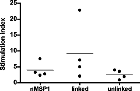
The outer membrane proteins (OMPs) of bacterial pathogens are essential for their growth and survival and especially for attachment and invasion of host cells. Since the outer membrane is the interface between the bacterium and the host cell, outer membranes and individual OMPs are targeted for development of vaccines against many bacterial diseases. Whole outer membrane fractions often protect against disease, and this protection cannot be fully reproduced by using individual OMPs. Exactly how the interactions among individual OMPs influence immunity is not well understood. We hypothesized that one OMP rich in T-cell epitopes can act as a carrier for an associated OMP which is poor in T-cell epitopes to generate T-dependent antibody responses, similar to the hapten-carrier effect. Major surface protein 1a (MSP1a) and MSP1b1 occur as naturally complexed OMPs in the Anaplasma marginale outer membrane. Previous studies demonstrated that immunization with the native MSP1 heteromer induced strong immunoglobulin G (IgG) responses to both proteins, but only MSP1a stimulated strong CD4+ T-cell responses. Therefore, to test our hypothesis, constructs of CD4+ T-cell epitopes from MSP1a linked to MSP1b1 were compared with individually administered MSP1a and MSP1b1 for induction of MSP1b-specific IgG. By linking the T-cell epitopes from MSP1a to MSP1b1, significantly higher IgG titers against MSP1b1 were induced. Understanding how the naturally occurring intermolecular interactions between OMPs influence the immune response may lead to more effective vaccine design.


Figures

FIG. 1.
FIG. 1.
Constructs used for immunization. Recombinant proteins contained epitopes used for purification at the N terminus (six-His tag) and the C terminus (FLAG tag) of the recombinant proteins. The R region is the repeat region of MSP1a; the F1, F2, and F3 regions are the areas in MSP1a used to map T-cell epitopes, which localize to F2 and F3 (6). (A) Recombinant T-cell epitope protein containing the F2-F3 region of MSP1a. (B) Recombinant full-length MSP1b1. (C) Recombinant linked protein containing the F2-F3 region of MSP1a linked to full-length MSP1b1. (D) Native MSP1 (nMSP1) consisting of naturally complexed full-length MSP1a and full-length MSP1b1. Open boxes indicate MSP1a sequences, black boxes indicate MSP1b1 sequences, and gray boxes indicate His and FLAG epitopes.
FIG. 2.
FIG. 2.
Isolation and detection of native MSP1 complex. (A) Purified native MSP1 complex separated on a 4 to 20% gradient SDS-PAGE gel and stained with Coomassie blue. Lane M, molecular size markers (molecular masses [in kDa] are indicated on the left); lane 1, 23 μg purified native MSP1 complex. (B) Detection of native MSP1 complex with MSP-specific MAbs. Lanes 1, 4, 7, 10, and 13, 0.1 μg of native MSP1 complex; lanes 2, 5, 8, 11, and 14, 10 μg of purified A. marginale strain St. Maries outer membranes; lanes 3, 6, 9, 12, and 15, 0.5 μg of uninfected erythrocyte membranes. The sizes (in kDa) of molecular size markers are indicated on the left. The blot was probed with MAb Ana22B1 specific for MSP1a (lanes 1 to 3), MAb AmR38A6 specific for MSP1b (lanes 4 to 6), MAb 115/362.17.19 specific for MSP2 (lanes 7 to 9), MAb 115/152.20.19 specific for MSP3 (lanes 10 to 12), and MAb AnaF16C1 specific for MSP5 (lanes 13 to 15).
FIG. 3.
FIG. 3.
Purification and detection of recombinant proteins. (A) One microgram of purified recombinant protein per lane separated on a 4 to 20% gradient SDS-PAGE gel and stained with Coomassie blue. Lane M, molecular size markers (molecular masses [in kDa] are indicated on the left); lane 1, recombinant T-cell epitopes; lane 2, recombinant MSP1b1; lane 3, recombinant linked protein. Asterisks indicate the position of the full-length protein. (B) Detection of recombinant proteins. Lanes 1 and 4, recombinant T-cell epitopes; lanes 2 and 5, recombinant MSP1b1; lanes 3 and 6, recombinant linked protein. Lanes 1 to 3 were incubated with a FLAG-specific MAb, and lanes 4 to 6 were incubated with an unrelated MAb specific for T. brucei.
FIG. 4.
FIG. 4.
MSP1b-specific IgG responses in immunized animals. Western blotting was performed to determine the serum titers specific for MSP1b1. Frozen and thawed sera were diluted 1:5,000 to 1:80,000. The IgG titers are expressed as the reciprocal of the lowest dilution at which a band was observed for MSP1b at 100 kDa. Each dot on the graph represents the serum titer for an individual, and the horizontal lines indicate the mean group titers. Significantly higher titers of MSP1b1-specific IgG were obtained for the group immunized with the linked construct than for the group immunized with the unlinked construct, as determined by the Wilcoxon ranked sum test (P = 0.031). nMSP1, native MSP1.
FIG. 5.
FIG. 5.
Proliferative responses of PBMC to recombinant MSP1b1. PBMC obtained 3 weeks after the fourth immunization were cultured for 6 days in triplicate with 0.01, 0.1, and 1 μg/ml recombinant MSP1b1 or recombinant OMP10 expressed from the same vector. To control for any proliferation due to contaminating E. coli products, the stimulation index was determined by dividing the mean cpm induced by MSP1b1 by the mean cpm induced by OMP10. The data for 1 μg/ml antigen are shown, and statistically significant differences were not detected between groups, as determined by ANOVA (P = 0.25).

Similar articles

Cited by

References

    1. Abbott, J. R., G. H. Palmer, K. A. Kegerreis, P. F. Hetrick, C. J. Howard, J. C. Hope, and W. C. Brown. 2005. Rapid and long-term disappearance of CD4+ T lymphocyte responses specific for Anaplasma marginale major surface protein-2 (MSP2) in MSP2 vaccinates following challenge with live A. marginale. J. Immunol. 1746702-6715. - PubMed
    1. Allred, D. R., T. C. McGuire, G. H. Palmer, S. R. Leib, T. M. Harkins, T. F. McElwain, and A. F. Barbet. 1990. Molecular basis for surface antigen size polymorphisms and conservation of a neutralization-sensitive epitope in Anaplasma marginale. Proc. Natl. Acad. Sci. USA 873220-3224. - PMC - PubMed
    1. Bavoil, P., A. Ohlin, and J. Schachter. 1984. Role of disulfide bonding in outer membrane structure and permeability in Chlamydia trachomatis. Infect. Immun. 44479-485. - PMC - PubMed
    1. Ben-Yedidia, T., and R. Arnon. 2006. Flagella as a platform for epitope-based vaccines. Isr. Med. Assoc. J. 8316-318. - PubMed
    1. Ben-Yedidia, T., and R. Arnon. 2005. Towards an epitope-based human vaccine for influenza. Hum. Vaccines 195-101. - PubMed

Publication types

MeSH terms

Substances