. 2007 Dec 24;204(13):3119-31.
doi: 10.1084/jem.20071724. Epub 2007 Dec 17.

The dermis contains langerin+ dendritic cells that develop and function independently of epidermal Langerhans cells

Affiliations

The dermis contains langerin+ dendritic cells that develop and function independently of epidermal Langerhans cells

Lionel Franz Poulin et al. J Exp Med. .

Abstract

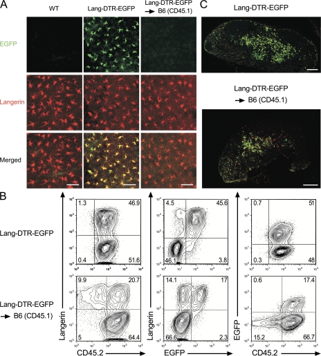
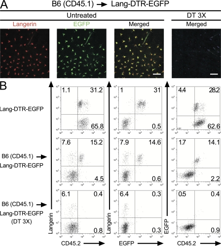
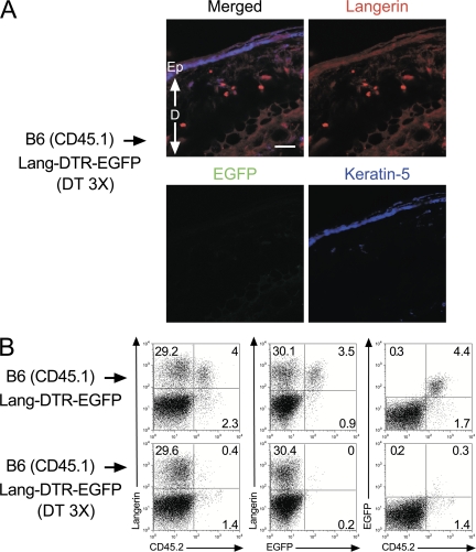
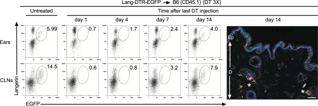
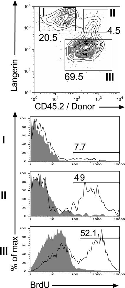
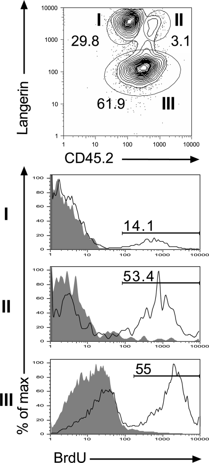
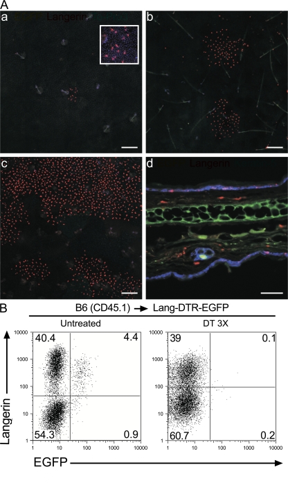
Langerhans cells (LCs) constitute a subset of dendritic cells (DCs) that express the lectin langerin and that reside in their immature state in epidermis. Paradoxically, in mice permitting diphtheria toxin (DT)-mediated ablation of LCs, epidermal LCs reappeared with kinetics that lagged behind that of their putative progeny found in lymph nodes (LNs). Using bone marrow (BM) chimeras, we showed that a major fraction of the langerin(+), skin-derived DCs found in LNs originates from a developmental pathway that is independent from that of epidermal LCs. This pathway, the existence of which was unexpected, originates in the dermis and gives rise to langerin(+) dermal DCs (DDCs) that should not be confused with epidermal LCs en route to LNs. It explains that after DT treatment, some langerin(+), skin-derived DCs reappear in LNs long before LC-derived DCs. Using CD45 expression and BrdU-labeling kinetics, both LCs and langerin(+) DDCs were found to coexist in wild-type mice. Moreover, DT-mediated ablation of epidermal LCs opened otherwise filled niches and permitted repopulation of adult noninflammatory epidermis with BM-derived LCs. Our results stress that the langerin(+) DC network is more complex than originally thought and have implications for the development of transcutaneous vaccines and the improvement of humanized mouse models.


Figures

Figure 1.
Figure 1.
Two developmental pathways give rise to langerin+ skin-derived DCs found in CLNs. (A) Staining of epidermal sheets from Lang-DTR-EGFP → B6 (CD45.1) chimeras, wild-type (WT), and Lang-DTR-EGFP mice with an anti-langerin antibody (red). As expected, the langerin+ cells found in the epidermis of Lang-DTR-EGFP mice differ from those of WT mice and of Lang-DTR-EGFP → B6 (CD45.1) chimeras in that they express EGFP. Genotypes are indicated for each column and markers on the left. Results are representative of at least 10 mice of each genotype. (B) Tissue-derived DCs found in CLNs were defined on the basis of their CD11cinter to high MHCIIhigh phenotype (Fig. S2), and analyzed for the expression of langerin versus CD45.2, langerin versus EGFP, and EGFP versus CD45.2. Contour plots from Lang-DTR-EGFP CLNs are also included for comparison. The percentages indicated for each window correspond to the percentage of cells among CD11cinter to high, MHCIIhigh DCs. Results are representative of at least five experiments. (C) Staining of a CLN section from a Lang-DTR-EGFP → B6 (CD45.1) chimera with an anti-langerin antibody (red) shows that host-derived (EGFP langerin+) LCs colocalize with donor-derived (EGFP+ langerin+) LCs. A stained section from Lang-DTR-EGFP mice is shown for comparison. Bars: (A) 50 μm; (C) 200 μm.
Figure 2.
Figure 2.
Donor-derived langerin+ DCs are found in skin explants. (A) Confocal microscopic analysis of epidermal sheets from B6 (CD45.1) → Lang-DTR-EGFP chimera that were left untreated (untreated) or had received 3 injections of DT (DT 3 X) a week apart were analyzed 1 d after the last DT injection. Staining with anti-langerin antibody (red) revealed the presence of langerin+ EGFP+ LCs in untreated sheets and their complete absence 1 d after the last DT injection. Bars: (untreated panel) 50 μm; (DT-treated panel) 300 μm. Results are representative of at least two experiments. (B) Ear explants from B6 (CD45.1) → Lang-DTR-EGFP chimera that had received 3 injections of DT (DT 3 X) were prepared 1 d after the last injection and cultured in the presence of CCL21. CD11c+ MHCII+ cells migrating out of the ear explants were analyzed by using langerin-CD45.2, langerin-EGFP, and EGFP-CD45.2 plots. Ear explants from untreated B6 (CD45.1) → Lang-DTR-EGFP chimera and Lang-DTR-EGFP mice were also included for comparison. The percentages indicated for each window corresponds to the percentage of cells among CD11c+, MHCII+ cells. Results are representative of at least two experiments.
Figure 3.
Figure 3.
Donor-derived langerin+ DCs are located in the skin dermis. (A) Staining of sections of ear from B6 (CD45.1) → Lang-DTR-EGFP chimera that had received 3 injections of DT and were prepared 1 d after the last injection were stained with anti-langerin (red) and anti–keratin 5 (blue) antibodies. Staining with the anti–keratin 5 antibody allowed visualization of the epidermis. Donor-derived, langerin+ DCs (EGFP, langerin+) are exclusively found in the dermis. Bar, 30 μm. Ep, epidermis; D, dermis. Results are representative of at least two experiments. (B) CLNs of DT-treated B6 (CD45.1) → Lang-DTR-EGFP chimera were collected 1 d after the last DT injection. Tissue-derived (CD11cinter to high, MHCIIhigh) DCs were analyzed as described in the legend of Fig. 1 for the expression of langerin-CD45.2, langerin-EGFP, and EGFP-CD45.2. The percentage indicated for each window corresponds to the percentage of cells among CD11cinter to high, MHCIIhigh DCs. Results are representative of at least 4 experiments.
Figure 4.
Figure 4.
Kinetics of reappearance of donor-derived langerin+ DCs after DT ablation. Ear explants and CLNs from Lang-DTR-EGFP → B6 (CD45.1) chimeras that had received 3 injections of DT 1 wk apart were prepared at the specified time points after the last DT treatment. For ears, analysis was performed on cells migrating out of CCL21-treated ear explants. Gated DCs (ears, CD11c+ MHCII+; CLNs, CD11cinter to high MHCIIhigh) were analyzed using langerin-EGFP plots. A window corresponding to the langerin+ EGFP+ subset is shown. The percentage indicated for this window corresponds to the percentage of donor-derived, langerin+, EGFP+ cells within the DC gates specified above. Also shown is a confocal microscopic analysis of a section of ear from a DT-treated Lang-DTR-EGFP → B6 (CD45.1) chimera 14 d after the last DT treatment. Staining with anti-langerin (red) and anti-keratin 5 (blue), revealed that donor-derived (EGFP+), langerin+ DCs are located exclusively in the dermis, whereas host-derived (EGFP), langerin+ DCs are found in the epidermis. Some donor-derived, langerin+ DDCs (marked with a star) are found adjacent to hair follicles. Results are representative of at least two experiments. Ep, epidermis; D, dermis. Bar, 30 μm.
Figure 5.
Figure 5.
BrdU incorporation of host- and donor-derived langerin+ DCs found in the skin. Lang-DTR-EGFP B6 (CD45.1) chimeras were exposed to BrdU for 1 wk, and ear explants were prepared. DCs migrating out of the ear explants were gated on CD11c+, MHCII+ cells and analyzed. Three subsets can be distinguished using langerin versus CD45.2 dot plots: host-derived LCs (subset I: langerin+, CD45.2, EGFP), donor-derived, langerin+ DDCs (subset II: langerin+, CD45.2+, EGFP+), and donor-derived, langerin DDCs (subset III: langerin, CD45.2+, EGFP). Histograms show BrdU incorporation for each subset. Gray-filled histograms correspond to control staining profile of mice that received no BrdU. Percentages of BrdU+ cells are indicated. Results are representative of at least two experiments.
Figure 6.
Figure 6.
Wild-type langerin+ DDCs express higher levels of CD45.2 than wild-type epidermal LCs. Wild-type C57BL/6 (CD45.2+) mice were left untreated or exposed to BrdU for 1 wk, and ear explants were prepared and cultured in the presence of CCL21. The MHCII+, CD11c+ DCs migrating out of the ear explants were analyzed using langerin-CD45.2 plots. Two discrete subsets can be readily discriminated among langerin+ DCs according to the levels of CD45.2 they express. Subset I corresponds to LCs and expresses intermediate levels of CD45.2, whereas subset II corresponds to langerin+ DDCs and expresses high levels of CD45.2. Subset III corresponds to langerin DDCs. Histograms show BrdU incorporation for each subset. Gray-filled histograms correspond to control staining profile of mice that received no BrdU. Percentages of BrdU+ cells are indicated. Results are representative of at least two experiments.
Figure 7.
Figure 7.
Donor-derived LCs colonize emptied epidermal niches. B6 (CD45.1) → Lang-DTR-EGFP chimeras that had received 3 injections of DT 1 wk apart were analyzed at various time points after the last injection. (A) Epidermal sheets 1 (a), 4 (b), and 7 wk (c) after the last DT injection were stained with anti-langerin antibody (red) and ToPro3 blue. Rare patches of donor-derived LCs (langerin+ EGFP) were found 1 wk after the last injection (arrow). An enlarged view (206.8 × 206.8 μm) of one of these patches is shown in the top right corner of a. d corresponds to a section of an ear from a DT-treated B6 (CD45.1) → Lang-DTR-EGFP chimera 7 wk after the last DT injection costained with anti-langerin (red) and –keratin 5 (blue) antibodies. Donor-derived LCs are found in both the dermis and epidermis. Bars: (a–c) 200 μm; (d) 50 μm. Results are representative of at least two experiments with two to three mice per experiment. Ep, epidermis; D, dermis. (B) Analysis of tissue-derived (CD11cinter to high, MHCIIhigh) DCs present in the CLNs from untreated and DT-treated B6 (CD45.1) → Lang-DTR-EGFP chimera. In the case of the DT-treated mice, CLNs were analyzed 7 wk after the last DT-injection. Dot plots show langerin-EGFP distribution. Consistent with the results shown in A, 7 wk after the last DT injection, the CLNs of DT-treated B6 (CD45.1) → Lang-DTR-EGFP chimera are deprived of host-derived LCs and contain an abundant population of donor-derived LCs. Results are representative of at least two experiments.

References

    1. Steinman, R.M., and M.C. Nussenzweig. 2002. Avoiding horror autotoxicus: the importance of dendritic cells in peripheral T cell tolerance. Proc. Natl. Acad. Sci. USA. 99:351–358. - PMC - PubMed
    1. Villadangos, J.A., and P. Schnorrer. 2007. Intrinsic and cooperative antigen-presenting functions of dendritic-cell subsets in vivo. Nat. Rev. Immunol. 7:543–555. - PubMed
    1. Larregina, A.T., and L.D. Falo. 2005. Changing paradigms in cutaneous immunology: adapting with dendritic cells. J. Invest. Dermatol. 124:1–12. - PubMed
    1. Carbone, F.R., G.T. Belz, and W.R. Heath. 2004. Transfer of antigen between migrating and lymph node-resident DCs in peripheral T-cell tolerance and immunity. Trends Immunol. 25:655–658. - PubMed
    1. Shortman, K., and S.H. Naik. 2007. Steady-state and inflammatory dendritic-cell development. Nat. Rev. Immunol. 7:19–30. - PubMed

Publication types

MeSH terms