. 2007 Dec 24;204(13):3147-56.
doi: 10.1084/jem.20071966. Epub 2007 Dec 17.

Identification of a novel population of Langerin+ dendritic cells

Affiliations

Identification of a novel population of Langerin+ dendritic cells

Laura S Bursch et al. J Exp Med. .

Abstract

Langerhans cells (LCs) are antigen-presenting cells that reside in the epidermis of the skin and traffic to lymph nodes (LNs). The general role of these cells in skin immune responses is not clear because distinct models of LC depletion resulted in opposite conclusions about their role in contact hypersensitivity (CHS) responses. While comparing these models, we discovered a novel population of LCs that resides in the dermis and does not represent migrating epidermal LCs, as previously thought. Unlike epidermal LCs, dermal Langerin(+) dendritic cells (DCs) were radiosensitive and displayed a distinct cell surface phenotype. Dermal Langerin(+) DCs migrate from the skin to the LNs after inflammation and in the steady state, and represent the majority of Langerin(+) DCs in skin draining LNs. Both epidermal and dermal Langerin(+) DCs were depleted by treatment with diphtheria toxin in Lang-DTREGFP knock-in mice. In contrast, transgenic hLang-DTA mice lack epidermal LCs, but have normal numbers of dermal Langerin(+) DCs. CHS responses were abrogated upon depletion of both epidermal and dermal LCs, but were unaffected in the absence of only epidermal LCs. This suggests that dermal LCs can mediate CHS and provides an explanation for previous differences observed in the two-model systems.


Figures
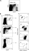
Figure 1.
Figure 1.
A novel population of radiosensitive LCs in dermis and skin draining LNs. Bone marrow chimeric mice were created by reconstituting lethally irradiated Lang-EGFP mice with bone marrow from congenic Lang-EGFP.SJL donors. (A) Representative flow cytometric analysis of LN CD11c+ I-Ab+ DCs. A broad I-Ab+ gate was used to exclude the small number of non-APCs that express CD11c. The plot shows distinct populations of EGFP−/int, CD8α+ DCs, and EGFPhi CD8α LCs. Only the latter will be referred to as LCs in this study. (B) Flow cytometric analysis of epidermal (a), dermal (d), and LN DCs (b and c) suspensions from Lang-EGFP.SJL into Lang-EGFP bone marrow chimeric mice. Plots on the right show CD45.1 (donor) and CD45.2 (host) expression on the indicated population. A prominent population of donor-derived LCs is observed in the LNs and dermis, but not in the epidermis.
Figure 2.
Figure 2.
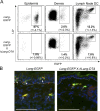
LCs are present in the dermis and skin draining LNs of hLang-DTA mice. (A) Flow cytometry analysis of I-Ab+ CD45.2+ cells in epidermal, dermal, and LN DCs suspensions from Lang-EGFP mice and Lang-EGFP x hLang-DTA Tg mice. In mice expressing the hLang-DTA transgene, EGFP+ cells are absent in epidermal cell suspensions, but present in both dermal and LN DCs preparations. Recovery of LCs in these tissues is ∼50–80% of that found in wild-type Lang-EGFP mice. The SD from two experiments with six Lang-EGFP and three Lang-EGFP x hLang-DTA mice is shown. (B) Immunofluorescence for Langerin and EGFP in cross sections of ear from Lang-EGFP mice and Lang-EGFP x hLang-DTA Tg mice reveals an absence of epidermal LCs (solid arrows) in Lang-EGFP x hLang-DTA Tg mice, whereas dermal Langerin+ DCs (line arrows) are present in both strains. Bar, 75 μm. Representative images from three Lang-EGFP and three Lang-EGFP x hLang-DTA mice are shown.
Figure 3.
Figure 3.
Donor-derived LCs display a distinct phenotype in dermis and skin draining LNs. Flow cytometry analysis of EGFP+ cells in epidermal, dermal, and LN DC suspensions from Lang-EGFP.SJL into Lang-EGFP bone marrow chimeric mice (A) or Lang-EGFP x hLang-DTA Tg mice (B). Host-derived epidermal DCs are predominantly Ep-CAM+ CD11b+, whereas donor-derived dermal DCs are predominantly CD11b Ep-CAM. Data shown are representative of 12 chimeras and 6 Lang-EGFP x hLang-DTA mice.
Figure 4.
Figure 4.
Dermal Langerin+ DCs express different levels of costimulatory and adhesion molecules. DCs were harvested from epidermis, dermis, lung, and skin draining LNs (SDLN) of 4 Lang-EGFP animals. CD11c+ populations were enriched from lung and LNs using MACS. Total EGFP+ cells are shown for epidermis and lung. In dermis and skin draining LNs, epidermal LCs and dermal Langerin+ DCs were distinguished using CD103 and CD11b as shown in Fig. S4. Isotype control staining is shown in the shaded histograms. Data are representative of two to six experiments. Fig. S4 is available at http://www.jem.org/cgi/content/full/jem.20071966/DC1.
Figure 5.
Figure 5.
Donor-derived LCs migrate from the skin to draining LNs. Lang-EGFP.SJL into Lang-EGFP bone marrow chimeras were painted with TRITC on the ear, and draining nodes were analyzed at 48, 72, or 96 h. (A) Dot plots show the fraction of TRITC+ CD11c+ DCs in draining (top) or nondraining (bottom) LNs after 72 h. (B) The histogram (left) shows Langerin (EGFP) expression on the migrated (TRITC+) DCs in the LNs at 72 h. The bar graph (right) shows the mean percentage of donor- (CD45.1+) or host-derived (CD45.2+) cells among migrated LCs (EGFP+) and other DCs (EGFP). Error bars indicate the SD. (C) Relative percentage of host- and donor-derived EGFP+ LCs in skin draining LNs over time (mean ± the SD) shown for three mice at each time point.
Figure 6.
Figure 6.
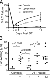
Dermal Langerin+ DCs contribute to the CHS response. (A) Time course of LC recovery in various tissues. Lang-DTREGFP mice were treated with 1 μg DT i.p. on day 0. The recovery of EGFP+ LCs in epidermis, dermis, and skin draining LNs was determined 2, 3, 4, 7, or 13 d later. The graph shows percentage of LC return, calculated as the percentage of EGFP+ DCs at the time of analysis compared with untreated control mice (mean± the SD) for three mice/group. (B) CHS. Lang-DTREGFP mice were treated with 1 μg DT i.p. at 1, 7, or 13 d before sensitization on the flank with DNFB. Mice were challenged with DNFB on the ear 5 d later, and ear swelling was measured 24 h after challenge. Positive control mice were not DT treated, and received both sensitization and challenge doses. Negative control mice were not sensitized, but received the challenge dose. Experimental results are shown from three independent experiments. The P value for specific comparisons is shown.

Similar articles

Cited by

References

    1. Stoitzner, P., K. Pfaller, H. Stossel, and N. Romani. 2002. A close-up view of migrating Langerhans cells in the skin. J. Invest. Dermatol. 118:117–125. - PubMed
    1. Stoitzner, P., S. Holzmann, A.D. McLellan, L. Ivarsson, H. Stossel, M. Kapp, U. Kammerer, P. Douillard, E. Kampgen, F. Koch, et al. 2003. Visualization and characterization of migratory Langerhans cells in murine skin and lymph nodes by antibodies against Langerin/CD207. J. Invest. Dermatol. 120:266–274. - PubMed
    1. Mayerova, D., E.A. Parke, L.S. Bursch, O.A. Odumade, and K.A. Hogquist. 2004. Langerhans cells activate naive self-antigen-specific CD8 T cells in the steady state. Immunity. 21:391–400. - PubMed
    1. Wolff, K. 1967. The fine structure of the Langerhans cell granule. J. Cell Biol. 35:468–473. - PMC - PubMed
    1. Valladeau, J., O. Ravel, C. Dezutter-Dambuyant, K. Moore, M. Kleijmeer, Y. Liu, V. Duvert-Frances, C. Vincent, D. Schmitt, J. Davoust, et al. 2000. Langerin, a novel C-type lectin specific to Langerhans cells, is an endocytic receptor that induces the formation of Birbeck granules. Immunity. 12:71–81. - PubMed

Publication types

MeSH terms