. 2008 Mar;28(5):1644-56.
doi: 10.1128/MCB.00325-07. Epub 2007 Dec 17.

Glucose-dependent insulinotropic polypeptide-mediated up-regulation of beta-cell antiapoptotic Bcl-2 gene expression is coordinated by cyclic AMP (cAMP) response element binding protein (CREB) and cAMP-responsive CREB coactivator 2

Affiliations

Glucose-dependent insulinotropic polypeptide-mediated up-regulation of beta-cell antiapoptotic Bcl-2 gene expression is coordinated by cyclic AMP (cAMP) response element binding protein (CREB) and cAMP-responsive CREB coactivator 2

Su-Jin Kim et al. Mol Cell Biol. 2008 Mar.

Abstract

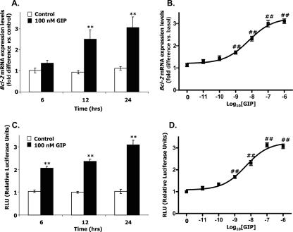
The cyclic AMP (cAMP)/protein kinase A (PKA) cascade plays a central role in beta-cell proliferation and apoptosis. Here, we show that the incretin hormone glucose-dependent insulinotropic polypeptide (GIP) stimulates expression of the antiapoptotic Bcl-2 gene in pancreatic beta cells through a pathway involving AMP-activated protein kinase (AMPK), cAMP-responsive CREB coactivator 2 (TORC2), and cAMP response element binding protein (CREB). Stimulation of beta-INS-1 (clone 832/13) cells with GIP resulted in increased Bcl-2 promoter activity. Analysis of the rat Bcl-2 promoter revealed two potential cAMP response elements, one of which (CRE-I [GTGACGTAC]) was shown, using mutagenesis and deletion analysis, to be functional. Subsequent studies established that GIP increased the nuclear localization of TORC2 and phosphorylation of CREB serine 133 through a pathway involving PKA activation and reduced AMPK phosphorylation. At the nuclear level, phospho-CREB and TORC2 were demonstrated to bind to CRE-I of the Bcl-2 promoter, and GIP treatment resulted in increases in their interaction. Furthermore, GIP-mediated cytoprotection was partially reversed by small interfering RNA-mediated reduction in BCL-2 or TORC2/CREB or by pharmacological activation of AMPK. The antiapoptotic effect of GIP in beta cells is therefore partially mediated through a novel mode of transcriptional regulation of Bcl-2 involving cAMP/PKA/AMPK-dependent regulation of CREB/TORC2 activity.


Figures

FIG. 1.
FIG. 1.
GIP increases the expression of Bcl-2 in INS-1 cells. (A) Time course of GIP-induced Bcl-2 expression levels. INS-1 (clone 832/13) cells were serum starved in 3 mM glucose-RPMI medium containing 0.1% BSA overnight, and GIP (100 nM) was added for the indicated periods of time. Total RNA was isolated from each sample, and real-time RT-PCR was performed to quantify Bcl-2 mRNA expression levels, shown as the relative difference from the control normalized to GAPDH expression levels. (B) Concentration response effect of GIP on Bcl-2 expression. INS-1 cells were treated as described above and treated with the indicated concentrations of GIP for 24 h. (C) Time course of GIP-induced Bcl-2 promoter activity. INS-1 cells were transfected with the −1714 Bcl-2 promoter reporter construct (2 μg) and pCMV β-galactosidase (1 μg) and serum starved in 3 mM glucose-RPMI medium containing 0.1% BSA overnight. Cells were then treated with 100 nM GIP for the indicated periods of time. The reporter activities are shown as the relative luciferase activity normalized to the β-galactosidase activity. (D) Concentration response effect of GIP on Bcl-2 promoter activity. INS-1 cells were transfected and treated as described above and incubated with the indicated concentrations of GIP for 24 h. All data represent three independent experiments, each carried out in triplicate. Significance was tested using ANOVA with a Newman-Keuls posthoc test (A and C) or Dunnet's multiple comparison test (B and D). **, P < 0.01 versus basal level; ##, P < 0.01 versus control.
FIG. 2.
FIG. 2.
GIP modulates CREB/TORC2 activity in INS-1 cells. (A) Time course of GIP-induced CREB phosphorylation. INS-1 cells were serum starved in 3 mM glucose-RPMI medium containing 0.1% BSA overnight and treated for the indicated periods of time with 100 nM GIP. Nuclear extracts were isolated, and Western blot analyses were performed as described in Materials and Methods, using antibodies against phospho-CREB and CREB. (B) Concentration-dependent effect of GIP on CREB phosphorylation. INS-1 cells were treated for 10 min with the indicated concentrations of GIP, and Western blot analyses were performed. (C) Time course of GIP-induced nuclear localization of TORC2. INS-1 cells were serum starved in 3 mM glucose-RPMI medium containing 0.1% BSA overnight and treated for the indicated periods of time with 100 nM GIP. Nuclear extracts were isolated, and Western blot analyses were performed as described in Materials and Methods using antibodies against TORC2. (D) Concentration response effect of GIP on nuclear localization of TORC2. INS-1 cells were treated for 30 min with the indicated concentrations of GIP, and Western blot analyses were performed. Western blots were quantified using densitometric analysis and are representative of three experiments. Significance was tested using ANOVA with Dunnet's multiple comparison test (**, P < 0.05 versus basal level). α, anti.
FIG. 3.
FIG. 3.
The cAMP/PKA pathway is involved in GIP-mediated CREB/TORC2 modulation and Bcl-2 activation. (A) Effect of PKA inhibition on GIP-induced CREB phosphorylation. INS-1 cells were treated as described in Materials and Methods and stimulated for 10 min with 100 nM GIP or 10 μM forskolin in the presence or absence of H-89. H-89 (10 μM) was added to cells during a 1-h preincubation as well as during GIP stimulation. Nuclear extracts were isolated from each sample, and Western blot analyses were performed. (B) Effect of PKA inhibition on GIP-mediated nuclear localization of TORC2. INS-1 cells were treated as described in Materials and Methods and stimulated for 30 min with 100 nM GIP or 10 μM forskolin in the presence or absence of H-89 (10 μM). Nuclear extracts were isolated from each sample, and Western blot analyses were performed. (C) Effect of PKA inhibition on GIP-induced Bcl-2 promoter activity. INS-1 cells were cotransfected with the −1714 Bcl-2 promoter reporter construct (2 μg) and pCMV β-galactosidase (1 μg) and serum starved in 3 mM glucose-RPMI medium containing 0.1% BSA overnight. Cells were then stimulated for 24 h with GIP (100 nM) or forskolin (10 μM) in the presence or absence of H-89. H-89 was present during a 1-h preincubation and GIP stimulation. The reporter activities are shown as the relative luciferase activity normalized to the β-galactosidase activity. All data represent three independent experiments, each carried out in triplicate. Significance was tested using ANOVA with a Newman-Keuls posthoc test. **, P < 0.05 versus basal level; ##, P < 0.05 versus GIP; and §§, P < 0.05 versus forskolin. α, anti.
FIG. 4.
FIG. 4.
GIP regulates the nuclear localization of TORC2 through a pathway involving activation of cAMP/PKA and decreased AMPK phosphorylation. (A) Concentration-dependent effect of GIP on phospho-AMPK (p-AMPK) Thr172. INS-1 cells were serum starved in 3 mM glucose-RPMI medium containing 0.1% BSA overnight and treated for 30 min with the indicated concentrations of GIP. Total cellular extracts were isolated, and Western blot analyses were performed using antibody against phospho-AMPK (Thr172), AMPK and β-actin. (B) Effect of AMPK modulation on TORC2 activity. INS-1 cells were treated for 30 min with GIP (100 nM) in the presence or absence of AMPK inhibitor (compound C; 40 μM) or AMPK activator (50 μM FPPF). AMPK inhibitor or activator was added to cells during a 1-h preincubation as well as during GIP stimulation. Nuclear or cytoplasmic extracts were isolated from each sample, and Western blot analyses were performed. TORC2 blots are from nuclear extracts, and phospho-AMPK Thr172 and β-actin blots are from cytoplasmic extracts. (C) Effect of PKA inhibition on GIP-mediated decreases in phospho-AMPK Thr172. INS-1 cells were treated as described above and stimulated for 30 min with GIP (100 nM) or forskolin (10 μM) in the presence or absence of H-89. H-89 (10 μM) was added to cells during a 1-h preincubation as well as GIP stimulation. Cytoplasmic extracts were isolated from each sample, and Western blot analyses were performed. Western blots are representative of three replicates.
FIG. 5.
FIG. 5.
GIP increases protein-protein interaction between phospho-CREB and TORC2 in the nucleus. (A) Coimmunoprecipitation. INS-1 cells were serum starved in 3 mM glucose-RPMI medium containing 0.1% BSA overnight and stimulated for 30 min with GIP (100 nM) or forskolin (10 μM) in the presence or absence of H-89 (10 μM). Nuclear extracts were isolated from each sample and immunoprecipitated (IP) with phospho-CREB (Ser133) followed by immunoblotting (IB) for TORC2. Input represents 1/10 of the total nuclear extract used in the Coimmunoprecipitation assay. (B) Confocal microscopy. INS-1 cells were treated as described above and fixed following stimulation with GIP (100 nM) or forskolin (10 μM) in the presence or absence of H-89 (10 μM). Immunocytochemical staining was performed using antibodies against phospho-CREB (Ser133; green) or TORC2 (red), and nuclei were stained with DAPI (blue). The scale bar indicates 20 μm, and all imaging data were analyzed using the Northern Eclipse program (version 6).
FIG. 6.
FIG. 6.
The rat Bcl-2 promoter contains a functional CRE. (A) A functional CRE was localized to one main region of Bcl-2 promoter by transient transfection analysis of serially deleted constructs. A schematic diagram of the Bcl-2 serial deletion reporter constructs and their potential CREB binding regions is presented. INS-1 cells were transfected with various serial deletion reporter constructs (2 μg) and pCMV β-galactosidase (1 μg) and starved overnight. Cells were then treated with 100 nM GIP for 24 h, and luciferase assays were performed. The reporter activities are shown as the relative luciferase activity normalized to the β-galactosidase activity. (B) Mutations in the CRE-I affect the GIP responsiveness of the rat Bcl-2 promoter. The sequences of WT and mutant CRE-I constructs are presented. All sequences of mutant CRE-I constructs were identical to the WT construct except for the indicated mutated sequences (underlined) in CRE-I. INS-1 cells were transfected with the WT construct (−1714 Bcl-2 promoter reporter construct; 2 μg) and pCMV β-galactosidase (1 μg) or mutated CRE-I constructs (M1 to M5 CRE-I; 2 μg) and pCMV β-galactosidase (1 μg). Cells were starved overnight and treated with 100 nM GIP for 24 h. Reporter activity is shown as the relative luciferase activity normalized to the β-galactosidase activity. (C and D) Representative gel picture of a ChIP assay for the binding of phospho-CREB (C) and TORC-2 (D) in the rat Bcl-2 promoter. INS-1 cells were serum starved in 3 mM glucose-RPMI medium containing 0.1% BSA overnight and stimulated for 10 min with 100 nM GIP or 10 μM forskolin in the presence or absence of H-89. H-89 (10 μM) was added to cells during a 1-h preincubation as well as during GIP stimulation. Phospho-CREB and TORC2 were immunoprecipitated from intact chromatin isolated from INS-1 cells using, respectively, anti-phospho-CREB (Ser133) and anti-TORC2 antibody. Precipitated DNA fragments were analyzed by PCR using primers flanking the CRE-I site in the Bcl-2 promoter. An isotype-matched immunoglobulin G was used as a negative control and 1% input (PCR product of 1/100 of the total isolated DNA used in the ChIP assay) was used as a positive control. All data represent three independent experiments, each carried out in duplicate. Significance was tested using ANOVA with a Newman-Keuls posthoc test. **, P < 0.05 versus basal level. IgG, immunoglobulin G.
FIG. 7.
FIG. 7.
RNA interference-mediated suppression of CREB and TORC2 reduces Bcl-2 expression, and inhibition of BCL-2 expression results in attenuation of GIP-mediated cytoprotection. (A) Concentration-dependent effect of CREB siRNA on CREB/TORC2/BCL-2 expression. MIN6 β cells were transfected with a pool of three siRNAs for CREB and incubated for 72 h. Western blot analyses were performed as described in Materials and Methods, using antibody against phospho-CREB (serine 133), CREB, TORC2, β-actin, and BCL-2. (B) Concentration-dependent effect of TORC2 siRNA on CREB/TORC2/BCL-2 expression. MIN6 β cells were transfected with a pool of three siRNAs for TORC2, and Western blot analyses were performed. (C) Effects of CREB and TORC2 siRNAs on GIP-mediated cytoprotection. MIN6 β cells were transfected with a pool of three siRNAs each for CREB and TORC2 (100 nM for each) and treated with thapsigargin (1 μM) for 8 h in the presence or absence of GIP (100 nM) under serum-free conditions. Caspase-3 activity was determined as described in Materials and Methods. (D) Concentration-dependent effect of BCL-2 siRNA on BCL-2 expression. MIN6 β cells were transfected with a pool of three siRNAs for BCL-2, and Western blot analyses were performed using antibodies against β-actin and BCL-2. Phospho-CREB, CREB, and TORC2 blots were performed with nuclear extracts, and BCL-2 and β-actin blots were performed with cytoplasmic extracts. (E) Effects of BCL-2 siRNA on GIP-mediated cytoprotection. MIN6 β-cells were transfected with a pool of three siRNAs for BCL-2 (200 nM) and treated with thapsigargin as described in panel C. All data represent three independent experiments, each carried out in duplicate, and Western blots are representative of three replicates. Significance was tested using ANOVA with a Newman-Keuls posthoc test. **, P < 0.05 versus control siRNA; ##, P < 0.05 versus control siRNA under apoptotic conditions; ^, P < 0.05 versus CREB plus TORC2 siRNAs under apoptotic conditions; §§, P < 0.05 versus BCL-2 siRNA under apoptotic conditions.
FIG. 8.
FIG. 8.
AMPK/TORC2/BCL-2 are involved in GIP-mediated β-cell survival. (A) Effects of AMPK/TORC2 modulation on GIP-mediated cytoprotection. MIN6 β cells were transfected with a pool of three siRNAs for TORC2 (200 nM) or control siRNAs (200 nM) and incubated in the presence or absence of AMPK inhibitor (40 μM), AMPK activator (50 μM), and/or GIP (100 nM) under apoptotic conditions (with 1 μM thapsigargin for 8 h; serum free). Caspase-3 activity was determined as described in Materials and Methods. (B) Effects of AMPK/TORC2 modulation on GIP-mediated trans activation of the Bcl-2 promoter. MIN6 β cells were transfected with a pool of three siRNAs for TORC2 (200 nM) and the −1714 Bcl-2 promoter reporter construct, and Bcl-2 promoter activity assays were performed in the presence or absence of AMPK inhibitor or activator with the treatment of GIP (100 nM) or forskolin (10 μM). All data represent three independent experiments, each carried out in duplicate. In panel A, control data are identical to those shown in Fig. 7D for the comparison. Significance was tested using ANOVA with a Newman-Keuls posthoc test. **, P < 0.05 versus control siRNA; ##, P < 0.05 versus control siRNA under apoptotic conditions; §§, P < 0.05 versus control siRNA under apoptotic conditions plus AMPK activator; ^, P < 0.05 versus TORC2 siRNA under apoptotic conditions; &&, P < 0.05 versus TORC2 siRNA under apoptotic conditions plus AMPK activator; ++, P < 0.05 versus TORC2 siRNA; and ••, P < 0.05 versus control siRNA plus AMPK activator.
FIG. 9.
FIG. 9.
Proposed pathway by which GIP increases Bcl-2 transcription in pancreatic β cells. GIP binding to the G protein-coupled GIP receptor (GIPR) activates a stimulatory G protein (Gs), resulting in stimulation of adenylate cyclase (AC) and accumulation of cAMP. Active PKA catalytic subunits (C), released following cAMP binding to PKA regulatory subunits, enter the nucleus and phosphorylate CREB (white arrow). An associated decrease in phosphorylation and activity of AMPK is thought to result in dephosphorylation of TORC2, thus allowing increased translocation from the cytoplasm into the nucleus. Dashed lines indicate inhibitory pathways mediated by AMPK phosphorylation of TORC2, resulting in cytoplasmic retention, and PKA inhibition of AMPK, allowing TORC2 dephosphorylation. TORC2 complexes with phospho-CREB Ser133 in the nucleus and binds to CRE-I of the rat Bcl-2 promoter, thus turning on the transcriptional machinery for up-regulation of Bcl-2.

Similar articles

Cited by

References

    1. Bonner-Weir, S. 2001. Beta-cell turnover: its assessment and implications. Diabetes 50(Suppl. 1)S20-S24. - PubMed
    1. Bonner-Weir, S., D. Deery, J. L. Leahy, and G. C. Weir. 1989. Compensatory growth of pancreatic beta-cells in adult rats after short-term glucose infusion. Diabetes 3849-53. - PubMed
    1. Brelje, T. C., J. A. Parsons, and R. L. Sorenson. 1994. Regulation of islet beta-cell proliferation by prolactin in rat islets. Diabetes 43263-273. - PubMed
    1. Buteau, J., W. El-Assaad, C. J. Rhodes, L. Rosenberg, E. Joly, and M. Prentki. 2004. Glucagon-like peptide-1 prevents beta cell glucolipotoxicity. Diabetologia 47806-815. - PubMed
    1. Butler, A. E., J. Janson, S. Bonner-Weir, R. Ritzel, R. A. Rizza, and P. C. Butler. 2003. β-Cell deficit and increased β-cell apoptosis in humans with type 2 diabetes. Diabetes 52102-110. - PubMed

Publication types

MeSH terms