. 2008 Feb;28(4):1393-403.
doi: 10.1128/MCB.01733-07. Epub 2007 Dec 17.

Spn1 regulates the recruitment of Spt6 and the Swi/Snf complex during transcriptional activation by RNA polymerase II

Affiliations

Spn1 regulates the recruitment of Spt6 and the Swi/Snf complex during transcriptional activation by RNA polymerase II

Lei Zhang et al. Mol Cell Biol. 2008 Feb.

Abstract

We investigated the timing of the recruitment of Spn1 and its partner, Spt6, to the CYC1 gene. Like TATA binding protein and RNA polymerase II (RNAPII), Spn1 is constitutively recruited to the CYC1 promoter, although levels of transcription from this gene, which is regulated postrecruitment of RNAPII, are low. In contrast, Spt6 appears only after growth in conditions in which the gene is highly transcribed. Spn1 recruitment is via interaction with RNAPII, since an spn1 mutant defective for interaction with RNAPII is not targeted to the promoter, and Spn1 is necessary for Spt6 recruitment. Through a targeted genetic screen, strong and specific antagonizing interactions between SPN1 and genes encoding Swi/Snf subunits were identified. Like Spt6, Swi/Snf appears at CYC1 only after activation of the gene. However, Spt6 significantly precedes Swi/Snf occupancy at the promoter. In the absence of Spn1 recruitment, Swi/Snf is constitutively found at the promoter. These observations support a model whereby Spn1 negatively regulates RNAPII transcriptional activity by inhibiting recruitment of Swi/Snf to the CYC1 promoter, and this inhibition is abrogated by the Spn1-Spt6 interaction. These findings link Spn1 functions to the transition from an inactive to an actively transcribing RNAPII complex at a postrecruitment-regulated promoter.


Figures
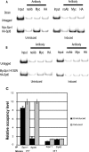
FIG. 1.
FIG. 1.
Spn1 and Spt6 are not coordinately recruited to CYC1, although Spt6 occupancy is Spn1 dependent. ChIP analysis using no antibody (noAb), anti-Myc antibody (Myc), or anti-HA antibody (HA) was performed on strains grown for 6 h under uninduced (2% dextrose) or induced (3% ethanol) conditions. (A) Spn1 continually occupies CYC1, while Spt6 occupies CYC1 only during activation. A control strain containing no tagged factors (untagged) was compared to a strain containing Myc-tagged Spn1 (Myc-Spn1) and HA-tagged Spt6 (HA-Spt6) via a representative ChIP assay. (B) Spn1 and Spt6 do not occupy CYC1 in the spn1-K192N background. A control strain containing no tagged factors (untagged) was compared to a strain containing Myc-tagged mutant Spn1-K192N (Myc-spn1-K192N) and HA-tagged Spt6 (HA-Spt6) via a representative ChIP assay. (C) Quantification of the relative Spn1 and Spt6 occupancy levels observed under uninduced and induced conditions. IP denotes the factor that was immunoprecipitated, whereas Strain indicates either the wild-type (WT) or spn1-K192N (MT) background. The protein occupancy level is represented as the ratio of signal from IP samples to that of the input minus background of a no-antibody control (n = 4; P < 0.005). Error bars indicate standard deviations.
FIG. 2.
FIG. 2.
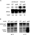
Different effects of SPT6 and SPN1 mutations on RNAPII transcription (A) Effect of spt6-1004 on the regulation of CYC1 activation. Total RNA from wild-type (WT) and spt6 mutant strains grown under partially repressed (in medium containing 2% dextrose) and activated (in medium containing 3% ethanol) conditions were analyzed by S1 nuclease assay using 32P-labeled CYC1 and tryptophan tRNA probes. tRNAw signal was used as a loading control to normalize the signal of CYC1 transcripts. A representative gel is shown. For quantification, the transcription level of CYC1 in the wild-type strain was set to 100%, and the values for the spt6 mutant strain from three independent experiments (±4%) are indicated. (B) Effect of an spn1 mutation on FLO8 transcription. Total RNA from wild-type and spn1 or spt6 mutant strains, grown at 30°C or after an 80-minute shift to 37°C, was subjected to Northern blot analysis for FLO8 RNA. The position of the transcript generated from the cryptic TATA element is indicated.
FIG. 3.
FIG. 3.
Spn1 associates with the RNAPII complex, whereas Spn1-K192N is diminished for this interaction. Protein extracts from strains were immunoprecipitated (IP) with protein A-Sepharose beads coupled with antibodies as indicated. Proteins of interest were detected by using corresponding antibodies or epitope tags and immunoblot analyses. Immunoprecipitation with anti-Spn1 antibodies (α-Spn1) pulled down RNAPII, and conversely, immunoprecipitation with antibodies to Rpb1, the largest subunit of RNAPII (α-RNAPII), pulled down Spn1. However, in the spn1-K192N mutant background, RNAPII association is significantly reduced with Spn1-K192N. Loads represent 10% of the input material for the IP (100% was loaded).
FIG. 4.
FIG. 4.
Determination of the occupancy levels of additional initiation complex components at CYC1. ChIP analysis was performed using antibodies for Rpb1 (RNAPII), the serine 5 phosphorylation-specific antibody (Ser5-P), or Myc antibodies and the indicated Myc-tagged strains for helicase subunits of TFIIH (Rad3 and Ssl2) or a capping enzyme subunit (Ceg1), under uninduced and induced conditions. The protein occupancy level is represented as the ratio of signal from immunoprecipitation samples to that of the input minus background control (irrelevant antibodies for RNAPII and serine 5 phosphorylation and an untagged strain for the remainder). In each case n = 3, with P < 0.005. Error bars indicate standard deviations.
FIG. 5.
FIG. 5.
SPN1 genetically interacts with SNF2, SNF5, SNF6, RTF1, and DST1. Yeast cells of the indicated strains were diluted serially and plated onto the indicated medium. Pictures were taken after 2 to 3 days of growth. The growth defects of the rtf1Δ and dst1Δ strains are exacerbated by the mutation in SPN1. The temperature-sensitive phenotype of the spn1 mutant (spn1-K192N) is suppressed by snf2Δ, snf5Δ, and snf6Δ, and the growth defects of the Swi/Snf mutants are suppressed by spn1-K192N. WT, wild type; MT, mutant.
FIG. 6.
FIG. 6.
The Swi/Snf complex is required for full activation of the CYC1 gene. (A) Quantification of the effects of Swi/Snf mutants on CYC1 transcription. S1 nuclease assay results show the effects of the Swi/Snf complex on CYC1 activation. The indicated strains were grown under uninduced and induced conditions, and total RNA was isolated and analyzed via S1 nuclease assay. tRNAw signal was used as a loading control to normalize signals of CYC1 transcripts. The fold changes in induction were calculated by dividing the signals of CYC1 transcripts under inducing conditions by the amount observed under noninducing conditions. The bar graph shows fold changes (mean ± standard deviation; P < 0.005) of CYC1 levels from each strain of four separate experiments. (B) Spn1 levels were comparable in all strains tested. Protein extracts from the indicated strains were analyzed by Western blotting using polyclonal anti-Spn1 antibody. TBP expression levels were used as an internal control. WT, wild type; MT, mutant.
FIG. 7.
FIG. 7.
Occupancy of Swi/Snf on the CYC1 promoter during activation in wild-type and spn1K192N backgrounds. ChIP analysis was performed on strains, as indicated in Fig. 1. (A) Swi/Snf occupies CYC1 during activation in a wild-type background. (B) The Swi/Snf complex autonomously occupies CYC1 in the spn1-K192N background. (C) Quantification of the relative Swi/Snf occupancy levels under uninduced and induced conditions. The protein occupancy level is represented as the ratio of signal from immunoprecipitation (IP) samples to that of the input minus background of a no-antibody control (n = 4; P < 0.005). WT, wild type; MT, mutant. Error bars indicate standard deviations.
FIG. 8.
FIG. 8.
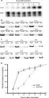
Time course of transcription and occupancy levels of Spt6 and Swi/Snf at CYC1. (A) S1 nuclease assay showing the time course of CYC1 activation. CYC1 is fully activated at 6 h of induction (medium containing 3% ethanol); tRNA was used as a loading control. (B) ChIP analysis showing the increase of Spt6 occupancy on the CYC1 promoter during 0 to 5 h of activation. Spt6 occupies the CYC1 promoter within 2 h after activation. (C) ChIP analysis showing the increase of the Swi/Snf complex occupancy on the CYC1 promoter during 0 to 5 h of activation. Swi/Snf occupancy parallels that of CYC1 transcription output. (D) Line graph showing the time course of Spt6 and Swi/Snf occupancy levels on the CYC1 gene upon activation. The levels of Spt6 and Swi/Snf occupancies at 6 h of activation were set as 100%. The occupancy levels of both factors at each time point were converted to the percentage of their maximum occupancy levels and graphed (n = 3; P < 0.005). Error bars indicate standard deviations.
FIG. 9.
FIG. 9.
A model for CYC1 gene regulation. (A) Under uninduced conditions, Spn1, TBP, RNAPII, TFIIH, and the capping enzyme subunit, Ceg1, are constitutively recruited to the CYC1 gene. In addition, serine 5 of the CTD of Rpb1 is phosphorylated. Spn1 occupancy prevents Swi/Snf interaction with the CYC1 promoter. (B) Under inducing conditions, Spt6 is recruited to the CYC1 promoter via interaction with Spn1. (C) Spt6 recruitment is followed by the recruitment of the Swi/Snf complex, which correlates with induced levels of gene expression (arrow).

Similar articles

Cited by

References

    1. Adelman, K., M. T. Marr, J. Werner, A. Saunders, Z. Ni, E. D. Andrulis, and J. T. Lis. 2005. Efficient release from promoter-proximal stall sites requires transcript cleavage factor TFIIS. Mol. Cell 17103-112. - PubMed
    1. Adkins, M. W., and J. K. Tyler. 2006. Transcriptional activators are dispensable for transcription in the absence of Spt6-mediated chromatin reassembly of promoter regions. Mol. Cell 21405-416. - PubMed
    1. Andrulis, E. D., E. Guzman, P. Doring, J. Werner, and J. T. Lis. 2000. High-resolution localization of Drosophila Spt5 and Spt6 at heat shock genes in vivo: roles in promoter proximal pausing and transcription elongation. Genes Dev. 142635-2649. - PMC - PubMed
    1. Awrey, D. E., N. Shimasaki, C. Koth, R. Weilbaecher, V. Olmsted, S. Kazanis, X. Shan, J. Arellano, C. H. Arrowsmith, C. M. Kane, and A. M. Edwards. 1998. Yeast transcript elongation factor (TFIIS), structure and function. II: RNA polymerase binding, transcript cleavage, and read-through. J. Biol. Chem. 27322595-22605. - PubMed
    1. Awrey, D. E., R. G. Weilbaecher, S. A. Hemming, S. M. Orlicky, C. M. Kane, and A. M. Edwards. 1997. Transcription elongation through DNA arrest sites. A multistep process involving both RNA polymerase II subunit RPB9 and TFIIS. J. Biol. Chem. 27214747-14754. - PubMed

Publication types

MeSH terms