. 2008 Mar;28(5):1584-95.
doi: 10.1128/MCB.00421-07. Epub 2007 Dec 17.

Corepressor CtBP and nuclear speckle protein Pnn/DRS differentially modulate transcription and splicing of the E-cadherin gene

Affiliations

Corepressor CtBP and nuclear speckle protein Pnn/DRS differentially modulate transcription and splicing of the E-cadherin gene

Roman Alpatov et al. Mol Cell Biol. 2008 Mar.

Abstract

CtBP is a transcriptional corepressor with tumorigenic potential that targets the promoter of the tumor suppressor gene E-cadherin. Pnn/DRS (Pnn) is a "nuclear speckle"-associated protein involved in mRNA processing as well as transcriptional regulation of E-cadherin via its binding to CtBP. Here, we show that CtBP can recruit Pnn to CtBP-associated complexes, resulting in Pnn-dependent chromatin remodeling at the E-cadherin promoter. In addition, CtBP and Pnn can differentially modulate E-cadherin mRNA splicing, with polymerase II serving as an interface in this event. Therefore, the Pnn/CtBP functional interplay represents a novel mechanism linking the corepressor CtBP and Pnn to the transcription-coupled mRNA splicing of a major tumor suppressor gene. Our findings implicate the existence of the molecular switches involved in tumorigenesis, which coordinate promoter-specific events and mRNA processing, by serving as bridging elements between the regulatory complexes both at gene promoters and within the mRNA splicing machineries.


Figures

FIG. 1.
FIG. 1.
Pnn is recruited to CtBP-associated repressor complexes. (A) CtBP bridges silencing machinery and its target gene promoters. (C) Pnn may sequester CtBP from the CtBP-associated repressors. (B) Alternatively, Pnn can be recruited to the repressor complex via CtBP. (D and E) HEK293 cells were cotransfected with vectors expressing ZEB1-myc (D) or mSin3A-myc (E) and CtBP1-Flag in the absence (lanes 1 and 3) or presence (lanes 2 and 4) of hPnn-GFP. Immunoprecipitations (IP) were performed using Flag affinity agarose, followed by Western blotting using anti-Pnn-143, anti-myc, and anti-Flag antibodies. Exogenous hPnn-GFP is indicated by an arrowhead, and endogenous Pnn is indicated by an arrow. An increase in Pnn levels did not appreciably affect the amount of ZEB1 or mSin3A coprecipitated with CtBP (D and E, respectively). Exogenous and endogenous Pnn could be detected in CtBP-Flag immunoprecipitates, indicating that Pnn may be present in ZEB1/CtBP1 and mSin3A/CtBP1 complexes.
FIG. 2.
FIG. 2.
Pnn can be recruited to the ZEB1/CtBP and mSin3A/CtBP complexes via CtBP. HEK293 cells were cotransfected with ZEB1-myc (A) or mSin3A-myc (B) in the presence of the short hairpin-mediated RNAi (shRNAi) vector for CtBP1 (lanes 2 and 4) or GFP RNAi (lanes 1 and 3). Immunoprecipitations (IP) were performed using anti-myc agarose, followed by Western blotting using anti-Pnn-143 and anti-myc antibodies. The amounts of Pnn in ZEB1 and mSin3A precipitates were diminished in the presence of CtBP RNAi, indicating that the Pnn association with the ZEB1/CtBP and mSin3A/CtBP repressor complexes was CtBP dependent. The bottom panels of A and B illustrate the efficiencies of CtBP RNAi in each corresponding experiment.
FIG. 3.
FIG. 3.
Pnn is present at the E-cadherin promoter, where it modulates the transcriptional state of chromatin. HeLa Pnn-Flag-HA cells or control HeLa cells were subjected to ChIP utilizing anti-Flag agarose, or antibodies against CBP, Pol II, AcH3K9, and AcH3K14 ChIPs were amplified using real-time PCR (qChIP) with either E-cadherin promoter (A)- or GAPDH promoter (B)-specific primers Pnn enrichment was detected at the E-cadherin promoter in HeLa Pnn-Flag-HA cells compared to the GAPDH promoter in the ChIP experiments utilizing anti-Flag agarose (lanes 1). In addition, HeLa Pnn-Flag-HA cells exhibited enrichment for CBP (A, lane 2), Pol II (A, lane 3), acetylated histone 3K9 (A, lane 4), and acetylated histone 3K14 (A, lane 5) at the E-cadherin promoter compared to control HeLa cells. The same degree of enrichment was not observed in the case of the GAPDH promoter (B). In contrast, the presence of diMeH3K9, a marker for silenced chromatin, was diminished in HeLa Pnn-Flag-HA cells compared to control HeLa cells. qChIP data are expressed as enrichment relative to the IgG control antibody. The enrichment in the case of HeLa Pnn-Flag-HA cells was expressed relative to the enrichment in the case of control HeLa cells, which was assigned a value of 1. Bars represent standard deviations of data from duplicate experiments.
FIG. 4.
FIG. 4.
Pnn interacts with the transcriptionally competent pool of Pol II, which can be depleted by CtBP. (A) Coimmunoprecipitations from the HeLa nuclear extracts using anti-Pol II (N-20) antibody followed by Western blotting using N-20 and anti-Pnn-143 antibodies. Pnn and Pol II were found to be associated. (B) Nuclear extracts from the HeLa Pnn-Flag-HA cells were used for immunoprecipitation using anti-HA antibody followed by Western blotting for HA, the preinitiation form of Pol II (CTD), initiation-specific Pol II (Pol II P-Ser5), and elongation-specific Pol II (Pol II P-Ser2). Pnn was found to be associated with transcriptionally competent forms of Pol II but not with the preinitiation form of Pol II. (C) Western blotting of control HeLa or HeLa CtBP-HA nuclei resuspended in SDS buffer. N-20 antibody detected a lower amount of hyperphosphorylated Pol II (IIo) in HeLa CtBP Flag-HA nuclei. Amounts of initiation-specific Pol II and elongation-specific Pol II were also diminished in the nuclei of CtBP-expressing cells. Nuclear lamins served as a loading control. (D) CTD-GST and Pnn-GST were subjected to the lysate-depended kinase reaction using control HeLa or HeLa CtBP-Flag-HA nuclear lysates. CTD-GST (top) but not Pnn-GST (bottom) exhibited a less pronounced molecular weight shift in HeLa CtBP Flag-HA lysates due in part to diminished Ser-5 phosphorylation (middle). Quantitation of the band intensity was performed using Adobe Photoshop software. (E) CTD-GST pull-downs from the kinase reactions were screened for bound proteins (as described in Materials and Methods). There were fewer Pnn and SR proteins bound to CTD in HeLa CtBP Flag-HA extracts.
FIG. 5.
FIG. 5.
Pnn can modulate E-cadherin mRNA splicing efficiency. (A) HEK293 cells were cotransfected with E-cadherin[E-cadEx4-Ex5] (left) or SV40[E-cadEx4-Ex5] (right) splicing reporters and increasing amounts of Pnn. The splicing efficiency was calculated as a ratio of intensities of the amplicons corresponding to the spliced mRNA to the sum of unspliced and spliced mRNA (total mRNA). Increasing Pnn expression resulted in an increased ratio of spliced mRNA to total mRNA in the context of the E-cadherin promoter but not the SV40 promoter. (B) Transfection of the Pnn RNAi vector resulted in a decreased ratio of spliced mRNA to total mRNA in the context of the E-cadherin promoter but not the SV40 promoter. Bars represent standard deviations of data from three independent experiments. (C) Mouse ES cells carrying an insertion of the neomycin resistance cassette into intron 8 of Pnn (3f/3f cells) or wild-type ES cells (wt/wt) were utilized as the source of RNA for splicing assays using RT-PCR. Unspliced message was detected using primers against E-cadherin intron 2, and total spliced E-cadherin message was detected using primers spanning the region between exons 4 and 8 (top). Splicing efficiency was calculated as a ratio of the intensity of the intronic amplicon to that of total spliced E-cadherin message (bottom). The ratio in the case of wt/wt cells was assigned a value of 1. 3f/3f cells exhibited an increase in the ratio of the intronic amplicon to total E-cadherin message compared to wt/wt cells, suggesting that in 3f/3f cells, E-cadherin splicing is inhibited. Bars represent standard deviations of data from duplicate experiments. (D) HeLa Pnn-Flag-HA cells were subjected to ChIP utilizing anti-Flag agarose followed by qPCR using E-cadherin intragenic primers or GAPDH intragenic primers as a control. Pnn enrichment was detected in the E-cadherin intragenic region but not the GAPDH intragenic region. The enrichment was expressed relative to the IgG control antibody, which was assigned a value of 1. Bars represent standard deviations of data from three experiments.
FIG. 6.
FIG. 6.
CtBP can negatively modulate E-cadherin mRNA splicing and can promote CD44 variant exon inclusion. (A) Splicing reactions using the E-cadherin splicing reporter were performed in the presence of the CtBP-Flag vector. CtBP-Flag expression resulted in a decreased ratio of spliced mRNA to total mRNA. Histograms on the right represent the ratio of spliced mRNA to total mRNA obtained from three independent assays. Bars represent standard deviations. (B) RT-PCR analysis of CD44 variant exon inclusion utilizing HeLa or HeLa CtBP-Flag-HA cells. RT reactions were subjected to PCR using upstream primers specific for variant exons 4, 5, 6, 7, and 8 (v4, v5, v6, v7, and v8, respectively) and a downstream primer (c3′) that anneals to a constitutive downstream 3′ region. Primers that anneal to the constitutive 5′ and 3′ regions (c5′ and c3′, respectively) were used to detect a standard CD44 form. (C) PCRs were run on a polyacrylamide gel followed by the quantification of the PCR band intensity (bottom). The degree of variant exon inclusion was calculated as a ratio of the intensity of amplicons of the variant exons to that of the amplicons of the standard CD44 form. The ratio of each variable amplicon (v4, v5, v6, v7, and v8) to a standard CD44 form in case of control HeLa cells was assigned a value of 1. HeLa CtBP-Flag-HA cells displayed increased expression of CD44 variants containing variable exons v4 and v5 but not v6 to v8 compared to control HeLa cells (C). Histograms represent average values of the ratios of each variable exon (v4, v5, v6, v7, and v8) to a standard CD44 form obtained from three independent experiments. Bars reflect standard deviations of data from three independent experiments.
FIG. 7.
FIG. 7.
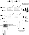
Diagram depicting a possible mechanism of Pnn and CtBP involvement in multiple steps of regulation of E-cadherin gene expression. (A) CtBP participates in the repression of the E-cadherin gene. (B) Pnn may be recruited to CtBP-associated repressor complexes via CtBP, where Pnn can attenuate CtBP-mediated repression. (C) CtBP and Pnn may also regulate E-cadherin gene expression at the mRNA level, where Pnn might couple transcription and splicing. Importantly, CtBP might modulate the degree of association of Pnn as well as other splicing factors with Pol II through the modulation of Pol II phosphorylation, thus affecting mRNA processing events.

Similar articles

Cited by

References

    1. Alpatov, R., G. C. Munguba, P. Caton, J. H. Joo, Y. Shi, M. E. Hunt, and S. P. Sugrue. 2004. Nuclear speckle-associated protein Pnn/DRS binds to the transcriptional corepressor CtBP and relieves CtBP-mediated repression of the E-cadherin gene. Mol. Cell. Biol. 2410223-10235. - PMC - PubMed
    1. Auboeuf, D., D. H. Dowhan, X. Li, K. Larkin, L. Ko, S. M. Berget, and B. W. O'Malley. 2004. CoAA, a nuclear receptor coactivator protein at the interface of transcriptional coactivation and RNA splicing. Mol. Cell. Biol. 24442-453. - PMC - PubMed
    1. Auboeuf, D., A. Honig, S. M. Berget, and B. W. O'Malley. 2002. Coordinate regulation of transcription and splicing by steroid receptor coregulators. Science 298416-419. - PubMed
    1. Balasubramanian, P., L. J. Zhao, and G. Chinnadurai. 2003. Nicotinamide adenine dinucleotide stimulates oligomerization, interaction with adenovirus E1A and an intrinsic dehydrogenase activity of CtBP. FEBS Lett. 537157-160. - PubMed
    1. Batsche, E., M. Yaniv, and C. Muchardt. 2006. The human SWI/SNF subunit Brm is a regulator of alternative splicing. Nat. Struct. Mol. Biol. 1322-29. - PubMed

Publication types

MeSH terms

LinkOut - more resources