. 2007 Dec 17;179(6):1193-204.
doi: 10.1083/jcb.200705180.

A molecular specificity code for the three mammalian KDEL receptors

Affiliations

A molecular specificity code for the three mammalian KDEL receptors

Irina Raykhel et al. J Cell Biol. .

Erratum in

  • J Cell Biol. 2008 Feb 11;180(3):645

Abstract

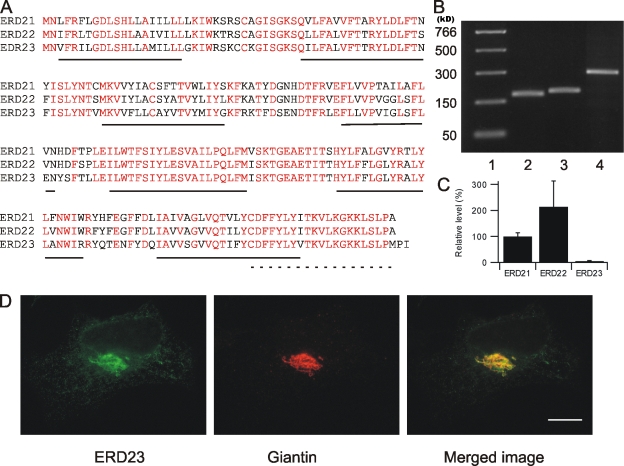
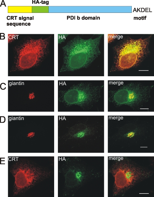
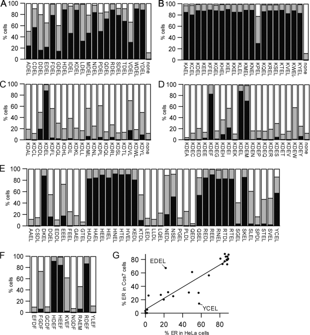
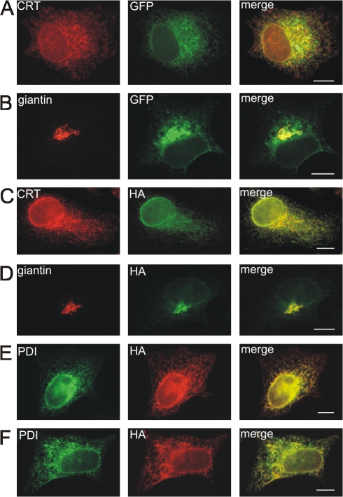
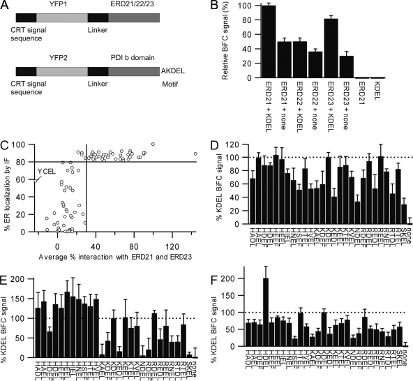
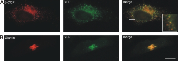
AC-terminal KDEL-like motif prevents secretion of soluble endoplasmic reticulum (ER)-resident proteins. This motif interacts with KDEL receptors localized in the intermediate compartment and Golgi apparatus. Such binding triggers retrieval back to the ER via a coat protein I-dependent pathway. To date, two human KDEL receptors have been reported. Here, we report the Golgi localization of a third human KDEL receptor. Using a reporter construct system from a screen of 152 variants, we identified 35 KDEL-like variants that result in efficient ER localization but do not match the current Prosite motif for ER localization ([KRHQSA]-[DENQ]-E-L). We cloned 16 human proteins with one of these motifs and all were found in the ER. A subsequent screen by bimolecular fluorescence complementation determined the specificities of the three human KDEL receptors. Each KDEL receptor has a unique pattern of motifs with which it interacts. This suggests a specificity in the retrieval of human proteins that contain different KDEL variants.


Figures

Figure 1.
Figure 1.
The three KDEL receptors found in human cells. (A) Alignment of the three human KDEL receptors. Identical amino acids for all three receptors are marked in red and the seven transmembrane regions are underlined by a continuous line. The commercial antibody against the human KDEL receptors was raised against the cytoplasmic C terminus of ERD21, which is underlined by a dotted line and shows 90% identity between the three receptors. (B) Total RNA was isolated from nontransfected HeLa cells. Three RT-PCR products of 178 (2), 190 (3), and 297 bp (4) represent the presence of the mRNA encoding for ERD21, 22, and 23 mRNA, respectively. The molecular mass markers (1) are indicated to the left of the agarose gel. (C) Relative quantification of mRNA for the three human receptors from HeLa cells by real-time PCR. The data represents the mean and standard deviation from 27 RT-PCR reactions normalized to the level of the mRNA for ERD21. (D) HeLa cells were transfected with the plasmid for ERD23 C-Myc tag, fixed, and double stained with antibodies against Myc and giantin. The merged image shows good colocalization of ERD23 and giantin. Bar, 10 μm.
Figure 2.
Figure 2.
The reporter construct used to determine the efficiency of ER localization based on KDEL motifs. (A) Schematic diagram of construct used. The reporter contains the b domain of PDI marked in blue, a HA tag marked in green, the signal sequence from CRT marked in yellow, and an ER-retention motif added to the C terminus. The reporter with the AKDEL- retention motif was used as a positive control and the reporter without a motif as a negative control. (B–E) HeLa cells were transfected with the plasmid for the HA-tagged reporter with the AKDEL motif added (B and C) or without the motif (D and E), fixed, and double stained with the antibodies against the HA tag and CRT (B and E) or giantin (C and D). Merged images show good colocalization of the reporter with the AKDEL motif with the ER marker (B) and the construct with no motif with the Golgi marker (D). Bars, 10 μm.
Figure 3.
Figure 3.
Immunofluorescence-based localization of the reporter constructs. For each construct, transient transfection of HeLa cells was performed in at least two independent experiments and the subcellular localization of the reporter construct was determined by examining at least 150 cells costained with a CRT antibody (ER marker) and a giantin antibody (Golgi marker). For each transfection, KDEL and no-motif controls were performed in parallel. Cells showing abnormal cell morphology or undergoing nuclear division were not included in the analysis. (A–F) The black bar represents the percentage of cells with ER-only localization, the gray bar the cells with mixed ER–Golgi localization, and the white bar the cells with Golgi-only localization. A, XDEL motifs; B, KXEL motifs; C, KDXL motifs; D, KDEX motifs; E, all other motifs found on human proteins ending L; F, other motifs found on human proteins ending F or M; G, linear correlation (R2 = 0.880) between HeLa and Cos results for 27 of the KDEL variants (note the marked YCEL and EDEL outliers).
Figure 4.
Figure 4.
Localization of tagged human proteins with and without their C-terminal retention motifs. HeLa cells were transfected with the plasmid for ERp18–GFP–LEDEL (A), ERp18–GFP–stop (B), GP7R–HA–KKEDL (C), GP7R–HA–stop (D), CRT3–HA–RRNEL (E), or CRT3–HA–stop (F), fixed, and stained with antibodies against CRT (A) or giantin (B) or double-stained with the antibodies against HA and CRT (C), HA and giantin (D), or HA and PDI (E and F). Merged images show good colocalization of the examined proteins with ER markers (A, C, E, and F) and Golgi markers (B and D). The examples shown are of ERp18 and GP7R changing localization and CRT3 staying in the ER after removing their putative ER-retention motif. Bars, 10 μm.
Figure 5.
Figure 5.
Analysis of the specificity of the three human KDEL receptors. (A) Schematic of the constructs used. (B) Proof of concept. For each of the three KDEL receptors the BiFC fluorescence signal counted from 3,000 cotransfected HeLa cells was significantly higher for the interaction with the KDEL-containing reporter than for the no-motif–containing reporter. The data represents the mean and standard deviation from four to six replicates. (C) Correlation between the ER localization of 85 of the KDEL variant reporter constructs and the mean BiFC signal for interactions with ERD21 and 23. A horizontal line is drawn at 80% ER localization (representing our definition of an efficient ER-localization motif) and a vertical line is drawn at 30% of the mean BiFC signal obtained with KDEL. Note the absence of motifs in the bottom right and the small number of motifs in the top left. (D–F) The BiFC measurements for each human KDEL receptor interacting with KDEL variants found on human proteins that result in efficient ER localization by immunofluorescence. The BiFC measurements are normalized to KDEL as 100% (dotted lines) and no motif as 0%. D, ERD21; E, ERD22; F, ERD23. The data represents the mean and standard deviation from six replicates.
Figure 6.
Figure 6.
Subcellular localization of BiFC interactions. HeLa cells were cotransfected with the plasmid for the Y1 fragment–tagged ERD21 (A and B) and the Y2 fragment–tagged AKDEL variant of the reporter construct (A) and the Y2 fragment–tagged no-motif reporter construct (B), fixed, and stained with antibodies against β-COP (A) or giantin (B). Merged images show good colocalization of the interacted proteins with the COP-I marker (A) and Golgi marker (B), respectively. The inset in A shows a closeup of the boxed area. Bars, 10 μm.

Similar articles

Cited by

References

    1. Alanen, H.I., K.E.H. Salo, M. Pekkala, H.M. Siekkinen, A. Pirneskoski, and L.W. Ruddock. 2003. a. Defining the domain boundaries of the human protein disulphide isomerases. Antioxid. Redox Signal. 5:367–377. - PubMed
    1. Alanen, H.I., R.A. Williamson, M.J. Howard, A.-K. Lappi, H.P. Jäntti, S.M. Rautio, S. Kellokumpu, and L.W. Ruddock. 2003. b. Functional characterization of ERp18, a new endoplasmic reticulum-located thioredoxin superfamily member. J. Biol. Chem. 278:28912–28920. - PubMed
    1. Alanen, H.I., R.A. Williamson, M.J. Howard, F.S. Hatahet, K.E.H. Salo, A. Kauppila, S. Kellokumpu, and L.W. Ruddock. 2006. ERp27, a new non-catalytic endoplasmic reticulum-located human protein disulfide isomerase family member, interacts with ERp57. J. Biol. Chem. 281:33727–33738. - PubMed
    1. Anelli, T., M. Alessio, A. Mezghrani, T. Simmen, F. Talamo, A. Bachi, and R. Sitia. 2002. ERp44, a novel endoplasmic reticulum folding assistant of the thioredoxin family. EMBO J. 21:835–844. - PMC - PubMed
    1. Anson, D.S., and J. Bielicki. 1999. Sulphamidase. Int. J. Biochem. Cell Biol. 31:363–367. - PubMed

Publication types