. 2007 Dec 17;179(6):1261-73.
doi: 10.1083/jcb.200703052.

Newly identified biologically active and proteolysis-resistant VEGF-A isoform VEGF111 is induced by genotoxic agents

Affiliations

Newly identified biologically active and proteolysis-resistant VEGF-A isoform VEGF111 is induced by genotoxic agents

Pierre Mineur et al. J Cell Biol. .

Abstract

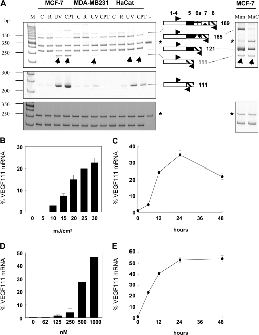
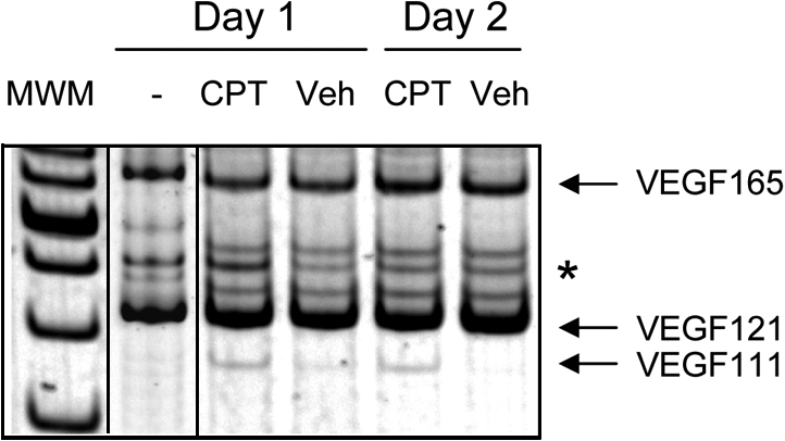
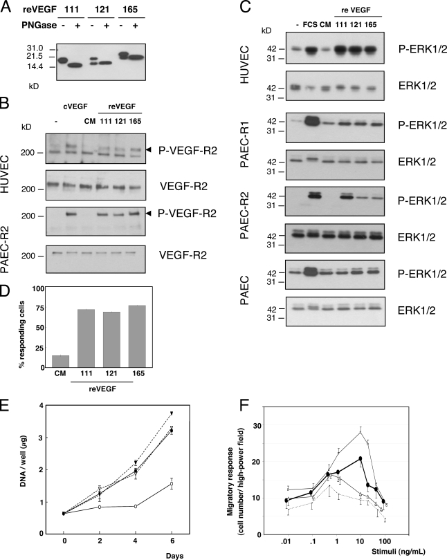
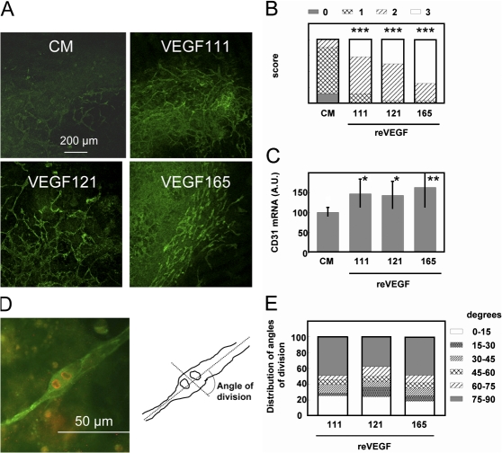
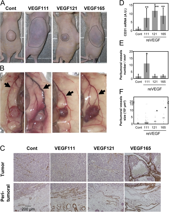
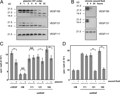
Ultraviolet B and genotoxic drugs induce the expression of a vascular endothelial growth factor A (VEGF-A) splice variant (VEGF111) encoded by exons 1-4 and 8 in many cultured cells. Although not detected in a series of normal human and mouse tissue, VEGF111 expression is induced in MCF-7 xenografts in nude mice upon treatment by camptothecin. The skipping of exons that contain proteolytic cleavage sites and extracellular matrix-binding domains makes VEGF111 diffusible and resistant to proteolysis. Recombinant VEGF111 activates VEGF receptor 2 (VEGF-R2) and extracellularly regulated kinase 1/2 in human umbilical vascular endothelial cells and porcine aortic endothelial cells expressing VEGF-R2. The mitogenic and chemotactic activity and VEGF111's ability to promote vascular network formation during embyonic stem cell differentiation are similar to those of VEGF121 and 165. Tumors in nude mice formed by HEK293 cells expressing VEGF111 develop a more widespread network of numerous small vessels in the peritumoral tissue than those expressing other isoforms. Its potent angiogenic activity and remarkable resistance to proteolysis makes VEGF111 a potential adverse factor during chemotherapy but a beneficial therapeutic tool for ischemic diseases.


Figures

Figure 1.
Figure 1.
Genotoxic agents induce the expression of VEGF111. The indicated cell lines were treated with 30 mJ/cm2 UV-B, 1 μM camptothecin, 5 mM l-mimosin, or 100 μg/ml mitomycin C for 24 h except otherwise indicated, and VEGF mRNA, VEGF111 mRNA, and 28S rRNA were measured by RT-PCR. (A) PAGE analysis of RT-PCR products of VEGF mRNA (top), VEGF111 mRNA (middle), and 28S rRNA (bottom). Arrows in the top panel indicate the VEGF111 amplification product. The drawings represent the exons (not to scale) encoding the corresponding VEGF isoforms. C, control cells; CPT, camptothecin treatment; M, 50-bp molecular weight markers; Mim, mimosin; MitC, mitomycin C; R, cells rinsed with red phenol–free medium but not irradiated; UV, UV-B irradiation; −, no cellular RNA. Asterisks indicate the RT-PCR products of synthetic RNA added to the test tubes to monitor reaction efficiency. (B–E) Dose-response analysis (B and D) and kinetics of induction (C and E) of the VEGF111 mRNA level in MCF-7 cells irradiated by UV-B (B and C) or treated with camptothecin (D and E). VEGF111 mRNA is given in percentages of the total VEGF isoform mRNA. Error bars represent SD.
Figure 2.
Figure 2.
Expression of VEGF111 in MCF-7 tumors treated with camptothecin. Estrogen-supplemented nude mice received a subcutaneous injection of MCF-7 cells (4 × 10⁶ cells) mixed with 100 μl matrigel in both flanks. After 3 wk, they received a daily intratumoral injection of 50 μl camptothecin (2 mg/ml) or the vehicle alone for 1 and 2 d. Tumors were collected 24 h after the last injection, and the VEGF mRNA expression was measured by RT-PCR. MWM, molecular weight marker; CPT, camptothecin treatment. The asterisk indicates the RT-PCR products of synthetic RNA added to the test tubes to monitor reaction efficiency.
Figure 3.
Figure 3.
VEGF111 is biologically active in vitro. (A) Expression of reVEGF in HEK293 cells and deglycosylation. HEK293 cells were transformed with vectors enabling the expression of VEGF111, 121, and 165. CM containing 20 ng VEGF as measured by ELISA was analyzed by Western blotting before and after treatment with N-glycosidase F (PNGase). (B and C) Serum-starved overnight HUVECs, PAEC, PAEC/R1, or PAEC/R2 (expressing VEGF-R1 or -R2) were treated with CM of HEK293 cells expressing VEGF111, 121, or 165 or with commercially available reVEGF165 (cVEGF) produced in bacteria (10 ng/ml each) or with 10% FCS for 5 min. Untreated cells (−) or cells treated with CM of control HEK293 cells (CM) served as controls. Total and phosphorylated VEGF-R2 (B) and ERK1/2 (C) were measured by Western blotting. (D) HUVECs labeled with Fluo3-AM were treated with CM of HEK293 cells expressing 10 ng/ml of VEGF111, 121, or 165 or CM from control HEK293 cells (CM). The percentage of cells responding by a 20% increase in intracellular free calcium concentration was recorded. (E) HUVECs were treated with HEK293 CM containing 10 ng/ml VEGF111 (black circles), 121 (open triangles), or 165 (black triangles) or CM from control HEK293 cells (open circles). DNA was measured in triplicate wells harvested as a function of time. (F) A modified Boyden chamber assay was used to measure the effect of VEGF111 (black circles), 121 (open triangles), 165 (open squares), and control medium (dotted line) on the chemotactic migration of PAEC/R2 cells. Error bars represent SD.
Figure 4.
Figure 4.
VEGF111 induces angiogenesis in embryoid bodies. Embryoid bodies were formed in the presence of CM from HEK293 cells expressing VEGF111, 121, or 165 or CM from control HEK293 cells (CM). (A) Representative microphotograph of embryoid bodies after immunofluorescent labeling of CD31. (B) Several microphotographs of six embryoid bodies from three independent experiments for each treatment were taken at random and analyzed by five investigators in double blind, and a score (from 0 to 3, where 0 indicates lack of vascular labeling and 3 indicates maximum labeling) was given to each sample. Statistical analysis was performed using a Chi square test. ***, P < 0.0001. (C) CD31 mRNA level was measured in embryoid bodies by RT-PCR. Statistical analysis was performed using a t test. *, P < 0.05; **, P < 0.01. (D and E) Embryoid bodies were differentiated in the presence of reVEGF111, 121, or 165. The angle between the plane of division of endothelial cells and the long axis of the vascular structures was analyzed after labeling of the vessels with anti-CD31 (green) and the mitotic cells with antiphosphohistone H3 (red) as illustrated by the cell in D for two sister cells having divided with a 90° angle. The angle of division plane was recorded for 43–63 cells in each group of embryoid bodies (n = 11). A scoring of the distribution of the angles is shown in E. Error bars represent SD.
Figure 5.
Figure 5.
VEGF111 induces angiogenesis in vivo. Mice were injected with a mixture of matrigel and HEK293 cells expressing reVEGF111, 121, or 165 or were transfected with empty vector (Cont) and killed after 3 wk. (A) Photographs of one representative mouse of each group before dissection. Dotted lines delimit the surface of the tumor and the peritumoral vascular network visible through the skin. (B) Photographs of one representative mouse of each group after dissection showing the tumor and the peritumoral tissue. Arrows indicate the lateral thoracic veins. (C) Histopathological analysis of the tumor and peritumoral tissues. Sections were stained with anti-αSMA antibodies and hematoxylin-eosin. (D) Expression of CD31 mRNA in the tumors: the mRNA was measured by RT-PCR in control HEK295 tumors (n = 21) and tumors grown from HEK293/111 (n = 6), HEK293/121 (n = 3), and HEK293/165 (n = 6). The data were corrected by the signals obtained for the 28S rRNA. (E) Number of vessels per millimeter squared of peritumoral tissue. The vessels were counted on paraffin sections of control mice (20 sections) and 111 (six sections), 121 (three sections), and 165 (six sections) mice. (F) Surface of the αSMA-positive vessels in the peritumoral tissue. Dots represent individual values, and horizontal bars indicate the mean value. *, P < 0.01; **, P < 0.0001 (versus control tumor). Statistical analysis was performed using the t test. Error bars represent SD.
Figure 6.
Figure 6.
VEGF111 is resistant to degradation by plasmin and proteases from nonhealing chronic wound fluid. (A and B) CM from HEK293 containing 20 ng reVEGF165, 121, and 111 were treated at 37°C with the indicated concentrations of plasmin for 4 h (A) or 10 μl of fluid collected from a chronic wound for the indicated times (B). The products were analyzed by Western blotting. (C and D) Cultures of HUVECs were treated with HEK293 cell CM containing 2 ng/ml VEGF111, 121, or 165, CM from control HEK293 (CM), or 2 ng/ml of commercial reVEGF165 (cVEGF) produced in bacteria either untreated or pretreated with 0.32 U plasmin for 4 h (C) or fluids collected from a chronic ulcer for 24 h (D). After 48 h, the cells were incubated with [3H]thymidine for 18 h, and TCA-precipitable radioactivity was measured. Statistics were performed using the analysis of variance test followed by the Tukey-Kramer test. *, P < 0.05; **, P < 0.001. Error bars represent SD.

Similar articles

Cited by

References

    1. Bates, D.O., T.G. Cui, J.M. Doughty, M. Winkler, M. Sugiono, J.D. Shields, D. Peat, D. Gillatt, and S.J. Harper. 2002. VEGF165b, an inhibitory splice variant of vascular endothelial growth factor, is down-regulated in renal cell carcinoma. Cancer Res. 62:4123–4131. - PubMed
    1. Blaudschun, R., C. Sunderkotter, P. Brenneisen, R. Hinrichs, T. Peters, L. Schneider, Z. Razi-Wolf, N. Hunzelmann, and K. Scharffetter-Kochanek. 2002. Vascular endothelial growth factor causally contributes to the angiogenic response upon ultraviolet B irradiation in vivo. Br. J. Dermatol. 146:581–587. - PubMed
    1. Brown, L.F., K.T. Yeo, B. Berse, T.K. Yeo, D.R. Senger, H.F. Dvorak, and L. van de Water. 1992. Expression of vascular permeability factor (vascular endothelial growth factor) by epidermal keratinocytes during wound healing. J. Exp. Med. 176:1375–1379. - PMC - PubMed
    1. Burchardt, M., T. Burchardt, A.G. Anastasiadis, R. Buttyan, A. de la Taille, A. Shabsigh, J. Frank, and R. Shabsigh. 2005. Application of angiogenic factors for therapy of erectile dysfunction: protein and DNA transfer of VEGF 165 into the rat penis. Urology. 66:665–670. - PubMed
    1. Caldwell, R.B., M. Bartoli, M.A. Behzadian, A.E. El-Remessy, M. Al-Shabrawey, D.H. Platt, G.I. Liou, and R.W. Caldwell. 2005. Vascular endothelial growth factor and diabetic retinopathy: role of oxidative stress. Curr. Drug Targets. 6:511–524. - PubMed

Publication types

MeSH terms