. 2008 Jan 29;98(2):399-409.
doi: 10.1038/sj.bjc.6604133. Epub 2007 Dec 18.

Antitumour effect of polyoxomolybdates: induction of apoptotic cell death and autophagy in in vitro and in vivo models

Affiliations

Antitumour effect of polyoxomolybdates: induction of apoptotic cell death and autophagy in in vitro and in vivo models

A Ogata et al. Br J Cancer. .

Abstract

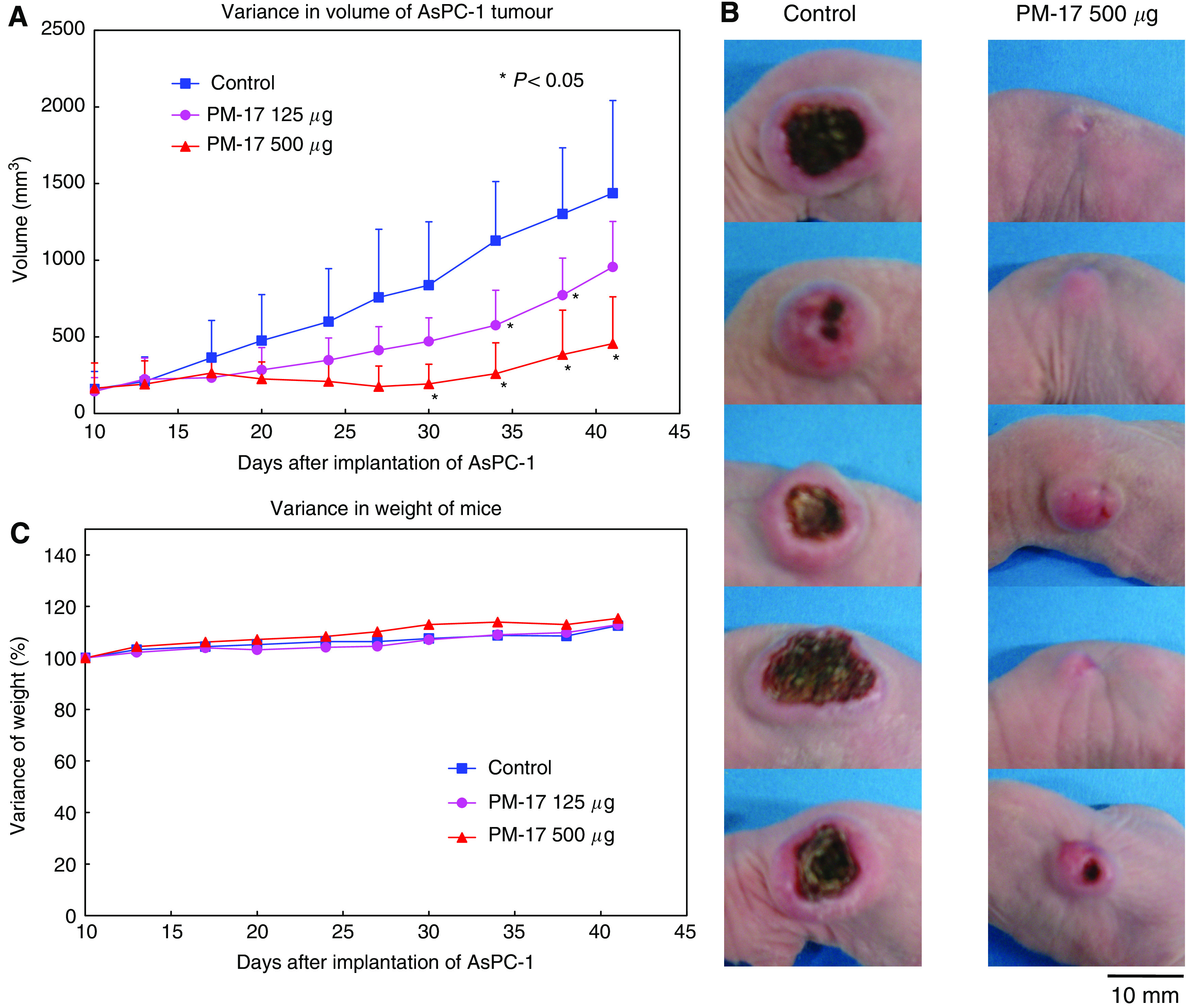
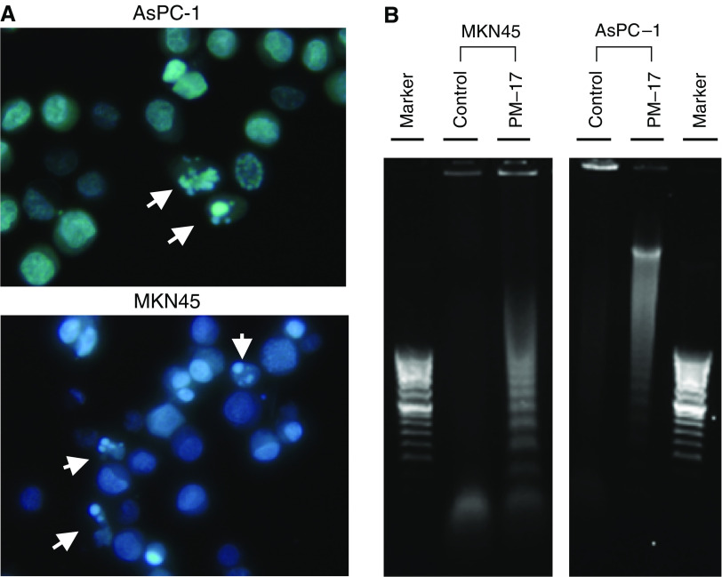
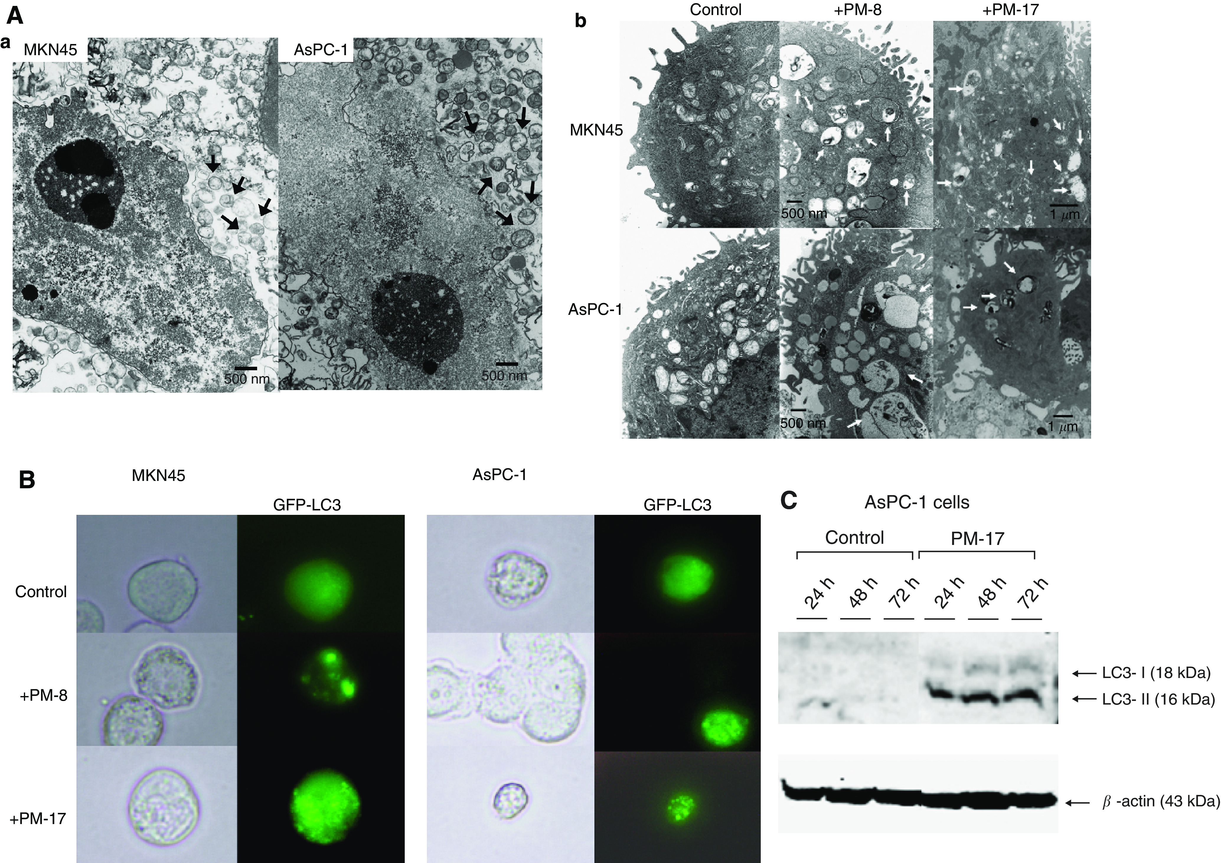
Polyoxomolybdates (PMs) as discrete molybdenum-oxide cluster anions have been investigated in the course of study of their medical applications. Here, we show the significant antitumour potency of the polyoxomolybdate [Me(3)NH](6)[H(2)Mo(V)(12)O(28)(OH)(12)(Mo(VI)O(3))(4)].2H(2)O (PM-17), which is a photo-reduced compound of [NH(3)Pr(i)](6)[Mo(7)O(24)].3H(2)O. The effect of PM-17 on the growth of cancer cell lines and xenografts was assessed by a cell viability test and analysis of tumour expansion rate. Morphological analysis was carried out by Hoechst staining, flow-cytometric analysis of Annexin V staining, terminal deoxynucleotidyl transferase-mediated 'nick-end' labelling staining, and electron-microscopic analysis. Activation of autophagy was detected by western blotting and fluorescence-microscopic analysis of the localisation of GFP-LC3 in transfected tumour cells. PM-17 inhibited the growth of human pancreatic cancer (AsPC-1) xenografts in a nude mice model, and induced morphological alterations in tumour cells. Correspondingly, PM-17 repressed the proliferation of AsPC-1 cells and human gastric cancer cells (MKN45) depending on the dose in vitro. We observed apoptotic patterns as the formation of apoptotic small bodies and translocation of phosphatidylserine by Hoechst staining and flow-cytometric analysis following Annexin V staining, and in parallel, autophagic conformation by the formulation of autophagosomes and localisation of GFP-LC3 by electron- and fluorescence-microscopic analysis.


Figures

Figure 1
Figure 1
PM-17 inhibits tumour growth depending on the dose in vivo. (A) Variance of tumour volume. After 10 days implantation of 2 × 10⁶ AsPC-1 cells in BALB/c nude mice, these mice were treated with 0.9% NaCl solution (▪, n=5), PM-17 (125 μg per 100 μl) (•, n=5), and PM-17 (500 μg per 100 μl) (▴, n=5) intratumorally. The intratumoral injections were performed for 10 days with 2 days’ intermission on day 6. The tumour sizes were measured using a micrometre caliper. Points, mean of tumour volume of five mice per group; bars, s.d. *P<0.05 compared with control. (B) Tumours of 41 days after implantation (left, saline control; right, PM-17 500 μg per body treated). (C) Alteration of weight of tumour-bearing mice. Points, mean of percentage of body weight per group.
Figure 2
Figure 2
The effect of PM-17 treatment on MKN45 cell and AsPC-1 cell survival in vitro. MKN45 cells were treated with PM-17 at the concentrations of 0–60 μg ml−1 for 24 h (A) and AsPC-1 cells were treated at the concentrations of 0–230 μg ml−1 for 24 h (B). Points, percentage of living cell in three independent experiments; bars, s.d.
Figure 3
Figure 3
Apoptosis detection in tumour cells treated with PM-17. (A) Hoechst 33342 staining. The tumour cells treated with PM-17 at the IC50 concentrations (AsPC-1, 175 μg ml−1; MKN45, 40 μg ml−1) for 24 h were harvested and stained with Hoechst 33342. Arrows show apoptotic small bodies. (B) DNA ladder formation of AsPC-1 and MKN45 after 24 h treatment without or with IC50 of PM-17. DNA fragments derived from nontreated cells and treated cells were electrophoresed in agarose gel with 100 bp DNA ladder marker. (C) Flow-cytometric analysis by double-staining of Annexin-V-FLUOS and PI on AsPC-1 and MKN45 after 24 and 48 h treatment without or with IC50 of PM-17. (D) Activation of apoptosis signalling via caspase-3 activation in MKN45 cells and AsPC-1 cells treated with PM-17. The variation of caspase-3 activity with time was analysed by measurement of chemiluminescence of a proluminescent caspase-3/7 substrate. Columns, means of luminescence intensity response to activity of caspase-3 in three independent experiments; bars, s.d. *P<0.05 compared with control, n=3. (E) TUNEL staining of tumours injected with PM-8 (4 mg per 100 μl) or PM-17 (500 μg per 100 μl) or 100 μl of 0.9% NaCl solution as control once for all. The observations were made with a wide or narrow field of view as shown in panels.
Figure 3
Figure 3
Apoptosis detection in tumour cells treated with PM-17. (A) Hoechst 33342 staining. The tumour cells treated with PM-17 at the IC50 concentrations (AsPC-1, 175 μg ml−1; MKN45, 40 μg ml−1) for 24 h were harvested and stained with Hoechst 33342. Arrows show apoptotic small bodies. (B) DNA ladder formation of AsPC-1 and MKN45 after 24 h treatment without or with IC50 of PM-17. DNA fragments derived from nontreated cells and treated cells were electrophoresed in agarose gel with 100 bp DNA ladder marker. (C) Flow-cytometric analysis by double-staining of Annexin-V-FLUOS and PI on AsPC-1 and MKN45 after 24 and 48 h treatment without or with IC50 of PM-17. (D) Activation of apoptosis signalling via caspase-3 activation in MKN45 cells and AsPC-1 cells treated with PM-17. The variation of caspase-3 activity with time was analysed by measurement of chemiluminescence of a proluminescent caspase-3/7 substrate. Columns, means of luminescence intensity response to activity of caspase-3 in three independent experiments; bars, s.d. *P<0.05 compared with control, n=3. (E) TUNEL staining of tumours injected with PM-8 (4 mg per 100 μl) or PM-17 (500 μg per 100 μl) or 100 μl of 0.9% NaCl solution as control once for all. The observations were made with a wide or narrow field of view as shown in panels.
Figure 4
Figure 4
Morphological analysis and detection of autophagic feature. MKN45 cells were treated with PM-8 (900 μg ml−1) or PM-17 (40 μg ml−1), and AsPC-1 cells were treated with PM-8 (1.65 mg ml−1) or PM-17 (175 μg ml−1). (A) Electron-microscopic analysis shows that mitochondria were disrupted. Inner constitution ((a), arrows) and appearance of quantity of autophagic vacuoles ((b), arrows) in MKN45 and AsPC-1 treated with IC50 of PM-8 or PM-17 for 24 h. (B) Subcellular localisation of GFP fusion LC3 protein in transfected MKN45 cells and AsPC-1 cells after treatment with or without IC50 of PM-8 or PM-17 for 24 h by fluorescent microscopy. (C) AsPC-1 cells treated with or without PM-17 (175 μg ml−1) for 24, 48, and 72 h. The cells were spun and lysed, and each 80 μg protein sample was loaded on SDS–PAGE gel and analysed by western blotting. Levels of LC3-I and -II expression were increased in cells treated with PM-17. Experiments were performed twice.

References

    1. Bernhard H, Jager-Arand E, Bernhard G, Heike M, Klein O, Riemann JF, Meyer zum Buschenfelde KH, Dippold W, Knuth A (1995) Treatment of advanced pancreatic cancer with 5-fluorouracil, folinic acid and interferon alpha-2A: results of a phase II trial. Br J Cancer 71: 102–105 - PMC - PubMed
    1. Boya P, Gonzalez-Polo RA, Casares N, Perfettini J, Dessen P, Larochette N, Métivier D, Meley D, Souquere S, Yoshimori T (2005) Inhibition of macroautophagy triggers apoptosis. Mol Cell Biol 25: 1025–1040 - PMC - PubMed
    1. Casper ES, Green MR, Kelsen DP, Heelan RT, Brown TD, Flombaum CD, Trochanowski B, Tarassoff PG (1994) Phase II trial of gemcitabine (2,2′-difluorodeoxycytidine) in patients with adenocarcinoma of the pancreas. Invest New Drugs 12: 29–34 - PubMed
    1. Crane CH, Abbruzzese JL, Evans DB, Wolff RA, Ballo MT, Delclos M, Milas L, Mason K, Charnsangavej C, Pisters PWT, Lee JE, Lenzi R, Vauthey JN, Wong ABS, Phan T, Nguyen Q, Janjan NA (2002) Is the therapeutic index better with gemcitabine-based chemoradiation than with 5-fluorouracil-based chemoradiation in locally advanced pancreatic cancer? Int J Radiat Oncol Biol Phys 52: 1293–1302 - PubMed
    1. Fakih MG (2006) Gemcitabine-induced rectus abdominus radiation recall. JOP 7: 306–310 - PubMed

Publication types