Review
. 2008 Jan;18(1):17-26.
doi: 10.1038/cr.2007.110.

DNA damage-induced cell death: lessons from the central nervous system

Affiliations
Review

DNA damage-induced cell death: lessons from the central nervous system

Helena Lobo Borges et al. Cell Res. 2008 Jan.

Abstract

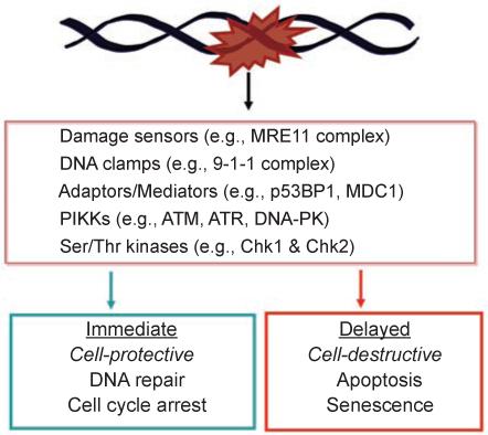
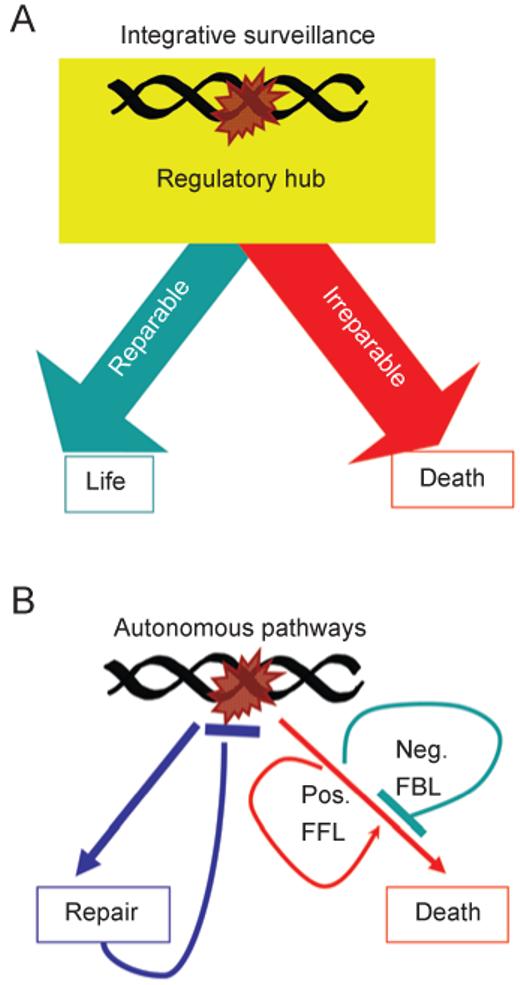
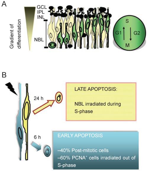
DNA damage can, but does not always, induce cell death. While several pathways linking DNA damage signals to mitochondria-dependent and -independent death machineries have been elucidated, the connectivity of these pathways is subject to regulation by multiple other factors that are not well understood. We have proposed two conceptual models to explain the delayed and variable cell death response to DNA damage: integrative surveillance versus autonomous pathways. In this review, we discuss how these two models may explain the in vivo regulation of cell death induced by ionizing radiation (IR) in the developing central nervous system, where the death response is regulated by radiation dose, cell cycle status and neuronal development.


Figures

Figure 1
Figure 1
DNA damage activates opposing biological outcomes through a common signaling network. Components of the DNA damage signal transduction network are listed in the box. Examples of proteins that sense DNA lesions, e.g., the Mre11 complex that detects strand breaks, and that assemble protein complexes, e.g., the 9-1-1 complex, p53BP1 and MDC1 are provided. The sensors, clamps and adaptors/mediators facilitate the activation of apical kinases belonging to the PIKK family, including ATM, ATR and DNA-PK. The PIKKs phosphorylate a large number of substrate proteins; among them are Chk1 and Chk2, which are serine/threonine kinases. The activation of DNA repair and cell cycle arrest is an immediate response to DNA damage, and these responses are designed to protect a damaged cell and to promote its recovery. The activation of apoptosis or senescence occurs later in time. These delayed responses are designed to eliminate a damaged cell. It is interesting to note that the DNA damage-signaling network can lead to two opposite biological outcomes.
Figure 2
Figure 2
Conceptual models for the coordination of DNA repair and cell death. (A) The “integrative surveillance” model: the DNA damage signaling network functions as a regulatory hub that makes calculated decisions, based on the status of DNA repair and the nature of DNA lesions to choose between life or death. (B) The “autonomous pathways” model: DNA damage activates DNA repair and cell death independently and automatically. The repair pathway extinguishes the damage signal. The death pathways (illustrated as a single entity to clarify the concept) contain intrinsic negative feedback loops (Neg. FBL) and positive feed-forward loops (Pos. FFL). These positive and negative regulatory loops are the causes of the delayed death response to DNA damage.
Figure 3
Figure 3
DNA damage-activated death machines. Summary of discussion in text on the various death mechanisms that have been shown to be activated by DNA damage in mammalian cell lines.
Figure 4
Figure 4
Role of the ATM-p53 pathway in IR-induced apoptosis of developing CNS. The phosphorylation sites are shown for human p53. (A) Summary of genetic studies that have identified players in IR-induced apoptosis in the developing CNS under variable doses (2, 5, 8, 10 and 14 Gy, see text for details). (B) Radiation dose affects the requirements for Atm and p53 in apoptosis. At 2 Gy, loss of Atm or p53, or serine-18 phosphorylation site in p53 (corresponding to human p53 serine-15 phosphorylation site), is sufficient to inactivate the apoptotic response to IR. While Atm through Chk2 can lead to p53 phosphorylation at Ser-23 (corresponding to human p53 serine-20), mutation at this site does not affect the apoptotic response in the developing retinas. With 2 Gy of IR, p53-heterozygosity is already showing defect in IR-induced apoptosis. Thus, IR-induced activation of ATM kinase and phosphorylation at serine-18 of a full complement of p53 protein from two wild-type alleles is required to induce apoptosis. At 14 Gy, Atm contributes to but is not required for apoptosis, whereas p53 is still required and p53 heterozygosity is sufficient to cause apoptosis. It thus appears that high-dose IR can super-activate p53 through Atm-dependent and independent (X) mechanisms such that half of the amount of p53 is sufficient to cause apoptosis.
Figure 5
Figure 5
Two waves of IR-induced apoptosis in the developing retina. (A) Cell layers in the developing retina of newborn mice or rats, and the interkinetic nuclear migration (cell cycle-related nuclear migration). The GCL and INL contain cells in an advanced stage of maturation. The IPL, inner plexiform layer, is a region of connections among cells. The NBL contains proliferating neuroblasts and recent post-mitotic cells. The gradient of differentiation is indicated on the left. Proliferating neuroblasts (white cells) and their nuclear migration along the phases of the cell cycle (G1, S, G2, M) are depicted. Black cells in the NBL are recent post-mitotic cells (G0 cells) that have exited the cell cycle and begun the process of terminal differentiation. (B) Two waves of apoptosis are induced by a single dose of IR as low as 0.5 Gy. Early apoptosis (6 h post-IR) involves recent post-mitotic cells (PCNA negative cells, also negative for differentiation markers), and proliferating neuroblasts not in S-phase at the time of irradiation (PCNA positive, BrdU negative). In this early wave, apoptosis correlates with an increase in lipid peroxidation and can be blocked by the anti-oxidant PDTC. Early apoptosis is also inhibited by blockage of protein synthesis (cycloheximide, CHX). The later wave of apoptosis (24 h post-IR) is comprised of neuroblasts that are in S-phase cells at the time of irradiation (PCNA positive, BrdU positive). Cells in the late wave of apoptosis are proliferating because they can be labeled with BrdU as late as 21.5 h post-IR. Because the late wave of apoptosis occurs in the S-phase zone, it suggests that apoptosis is triggered as cells re-enter the second S-phase after IR. However, we cannot rule out the possibility that these damaged cells remain in S-phase and continue to synthesize DNA for 24 h before dying to IR-induced damage. The later wave of apoptosis is not inhibited by the anti-oxidant PDTC but it is blocked by cycloheximide. Both waves of apoptosis require Atm and Serine-18 phosphorylation of p53 [48].

Similar articles

Cited by

References

    1. Rich T, Allen RL, Wyllie AH. Defying death after DNA damage. Nature. 2000;407:777–783. - PubMed
    1. Blank M, Shiloh Y. Programs for cell death: apoptosis is only one way to go. Cell Cycle. 2007;6:686–695. - PubMed
    1. Roos WP, Kaina B. DNA damage-induced cell death by apoptosis. Trends Mol Med. 2006;12:440–450. - PubMed
    1. Bentle MS, Bey EA, Dong Y, Reinicke KE, Boothman DA. New tricks for old drugs: the anticarcinogenic potential of DNA repair inhibitors. J Mol Histol. 2006;37:203–218. - PubMed
    1. Michod D, Widmann C. DNA-damage sensitizers: potential new therapeutical tools to improve chemotherapy. Crit Rev Oncol Hematol. 2007;63:160–171. - PubMed

Publication types