

Models of dengue virus infection
- PMID: 18087566
- PMCID: PMC1949394
- DOI: 10.1016/j.ddmod.2006.03.014
Models of dengue virus infection
Abstract
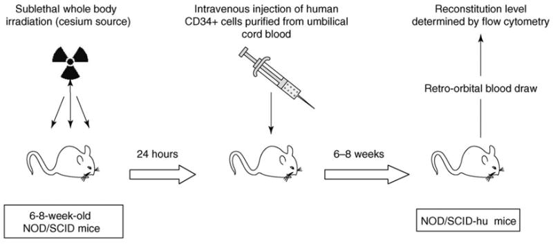
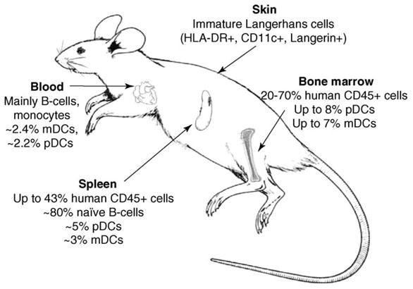
The need for models of dengue disease has reached a pinnacle as the transmission of this mosquito-borne virus has increased dramatically. Little is known about the mechanisms that lead to dengue fever and its more severe form, dengue hemorrhagic fever; this is owing to the fact that only humans show signs of disease. In the past 5 years, research has better identified the initial target cells of infection, and this has led to the development of models of infection in primary human cell cultures. Mouse-human chimeras, containing these target cells, have also led to progress in developing animal models. These advances should soon end the stalemate in testing antivirals and vaccine preparations that had necessarily been done in incomplete or irrelevant models.
Figures
References
-
- World Health Organization. Dengue/dengue hemorrhagic fever. Fact Sheet no. 117. 2002. http://www.who.int/mediacentre/factsheets/fs117/en/
-
- Halstead SB, et al. The future of dengue vaccines. Lancet. 2002;360:1243–1245. - PubMed
Grants and funding
LinkOut - more resources
Full Text Sources
Other Literature Sources
Miscellaneous