. 2008 May;105(3):605-16.
doi: 10.1111/j.1471-4159.2007.05155.x. Epub 2007 Dec 6.

The newly synthesized pool of dopamine determines the severity of methamphetamine-induced neurotoxicity

Affiliations

The newly synthesized pool of dopamine determines the severity of methamphetamine-induced neurotoxicity

David M Thomas et al. J Neurochem. 2008 May.

Abstract

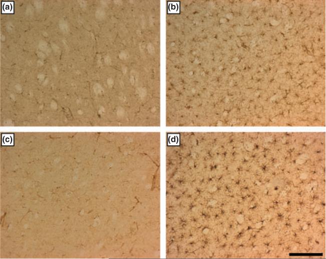
The neurotransmitter dopamine (DA) has long been implicated as a participant in the neurotoxicity caused by methamphetamine (METH), yet, its mechanism of action in this regard is not fully understood. Treatment of mice with the tyrosine hydroxylase (TH) inhibitor alpha-methyl-p-tyrosine (AMPT) lowers striatal cytoplasmic DA content by 55% and completely protects against METH-induced damage to DA nerve terminals. Reserpine, by disrupting vesicle amine storage, depletes striatal DA by more than 95% and accentuates METH-induced neurotoxicity. l-DOPA reverses the protective effect of AMPT against METH and enhances neurotoxicity in animals with intact TH. Inhibition of MAO-A by clorgyline increases pre-synaptic DA content and enhances METH striatal neurotoxicity. In all conditions of altered pre-synaptic DA homeostasis, increases or decreases in METH neurotoxicity paralleled changes in striatal microglial activation. Mice treated with AMPT, l-DOPA, or clorgyline + METH developed hyperthermia to the same extent as animals treated with METH alone, whereas mice treated with reserpine + METH were hypothermic, suggesting that the effects of alterations in cytoplasmic DA on METH neurotoxicity were not strictly mediated by changes in core body temperature. Taken together, the present data reinforce the notion that METH-induced release of DA from the newly synthesized pool of transmitter into the extracellular space plays an essential role in drug-induced striatal neurotoxicity and microglial activation. Subtle alterations in intracellular DA content can lead to significant enhancement of METH neurotoxicity. Our results also suggest that reactants derived from METH-induced oxidation of released DA may serve as neuronal signals that lead to microglial activation early in the neurotoxic process associated with METH.


Figures

Fig. 1
Fig. 1
Effects of AMPT and reserpine on DA depletion caused by a neurotoxic METH regimen. Mice (n = 5-8 per group) were treated with AMPT (4 × 100 mg/kg; t = -24, -16, -4 and -1 h) or reserpine (2.5 mg/kg; t = -24 h) alone, and before a neurotoxic METH regimen (4 × 5 mg/kg; t = 0, 2, 4, and 6 h). Striatal DA levels were determined at (a) t = 0, (b) t = 2 days and (c) t = 7 days. Results are presented as mean ± SEM relative to controls. Significant differences were determined via one-way ANOVA followed by Tukey's multiple comparison test, and are indicated as follows: *p < 0.01 relative to control (CON); #p < 0.01 relative to METH; ^ p < 0.05 relative to METH.
Fig. 2
Fig. 2
Effects of AMPT and reserpine on microglial activation caused by a neurotoxic METH regimen. Mice (n = 3-5 per group) were treated as described in Fig. 1 and analyzed for microglial activation in the striatum 2 days after the last METH injection. Microglia counts were obtained as described in the Materials and methods and are presented as means ± SEM. Treatment conditions and microglia counts for each panel are (a) Control (14 ± 1), (b) METH (142 ± 5), (c) AMPT + METH (18 ± 2) and (d) Reserpine + METH (179 ± 4). Significant differences were determined via oneway ANOVA followed by Tukey's multiple comparison test: p < 0.01, METH and Reserpine + METH relative to control; Reserpine + METH relative to METH. Scale bar represents 150 μm.
Fig. 3
Fig. 3
Effects of a neurotoxic METH regimen on core body temperatures of mice pre-treated with AMPT or reserpine. Mice (n = 5 per group) were treated as indicated, and core body temperatures monitored by telemetry every hour for 8 h. Results are presented as mean body temperature (°C) for each group at the indicated time points. METH was administered at t = 0, 120, 240, and 360 min. SEM bars were omitted for the sake of clarity and were < 10% of the mean in all groups. The reserpine group is the only statistically significant treatment effect overall (p < 0.0001, two-way ANOVA), and was also different from all other conditions (p < 0.01, Bonferroni's post-test).
Fig. 4
Fig. 4
Effect of L-DOPA on DA depletion caused by a neurotoxic METH regimen. L-DOPA was administered to (a) AMPT treated and (b) non-AMPT treated mice (n = 5-8 per group) to assess its effect on METH-induced DA depletion. Striatal DA levels were determined 2, 7, or 14 days after the METH regimen. Results are presented as mean ± SEM relative to controls. Significant differences were determined via one-way ANOVA followed by Tukey's multiple comparison test, and are indicated as follows: *p < 0.01 relative to control (CON); #p < 0.01 relative to METH.
Fig. 5
Fig. 5
Effects of L-DOPA on microglial activation caused by a neurotoxic METH regimen. Mice (n = 3-5 per group) were treated as described in Fig. 4 and analyzed for microglial activation in the striatum 2 days after the last METH injection. Microglia counts were obtained as described in the Materials and Methods and are presented as means ± SEM. Treatment conditions and microglia counts for each panel are (a) Control (17 ± 2), (b) METH (155 ± 10), (c) AMPT + L-DOPA + METH (171 ± 4) and (d) L-DOPA + METH (178 ± 3). Significant differences were determined via one-way ANOVA followed by Tukey's multiple comparison test: p < 0.01, all conditions relative to control; p < 0.05 L-DOPA + METH relative to METH. Scale bar represents 150 μm.
Fig. 6
Fig. 6
Effects of clorgyline on DA depletion caused by a neurotoxic METH regimen. Mice (n = 5-8 per group) were treated with clorgyline (10 mg/kg) alone, and 1 h before a neurotoxic METH regimen. Striatal DA levels were determined 2, 7, or 14 days after the METH regimen. Results are presented as mean ± SEM relative to controls. Significant differences were determined via one-way ANOVA followed by Tukey's multiple comparison test, and are indicated as follows: *p < 0.01 relative to control (CON); #p < 0.01 relative to METH.
Fig. 7
Fig. 7
Effects of clorgyline on microglial activation caused by a neurotoxic METH regimen. Mice (n = 3-5 per group) were treated as described in Fig. 6 and analyzed for microglial activation in the striatum 2 days after the last METH injection. Microglia counts were obtained as described in the Materials and Methods and are presented as means ± SEM. Treatment conditions and microglia counts for each panel are (a) control (15 ± 1), (b) METH (149 ± 5), (c) clorgyline (21 ± 2) and (d) clorgyline + METH (186 ± 9). Significant differences were determined via one-way ANOVA followed by Tukey's multiple comparison test: p < 0.01, METH and clorgyline + METH relative to control; p < 0.01, clorgyline + METH relative to METH. Scale bar represents 150 μm.
Fig. 8
Fig. 8
Effects of a neurotoxic METH regimen on core body temperatures of mice pre-treated with clorgyline or L-DOPA. Mice (n = 5 per group) were treated as indicated, and core body temperatures monitored by telemetry every hour for 8 h. Results are presented as mean body temperature (°C) for each group at the indicated time points. Clorgyline was administered at t = 0; METH was administered at t = 60, 180, 300 and 420 min. SEM bars were omitted for the sake of clarity and were < 10% of the mean in all groups. The clorgyline + METH group is the only statistically significant treatment effect overall (p < 0.001, two-way ANOVA), and is also different from control and clorgyline only conditions (p < 0.01, Bonferroni's post-test). Note that the ordinate scale used in this figure is expanded by comparison to Fig. 3 to make viewing of all body temperature traces easier.

References

    1. Albers DS, Sonsalla PK. Methamphetamine-induced hyperthermia and dopaminergic neurotoxicity in mice: pharmacological profile of protective and nonprotective agents. J. Pharmacol. Exp. Ther. 1995;275:1104–1114. - PubMed
    1. Benkovic SA, O'Callaghan JP, Miller DB. Sensitive indicators of injury reveal hippocampal damage in C57BL/6J mice treated with kainic acid in the absence of tonic-clonic seizures. Brain Res. 2004;1024:59–76. - PubMed
    1. Block ML, Zecca L, Hong JS. Microglia-mediated neurotoxicity: uncovering the molecular mechanisms. Nat Rev Neurosci. 2007;8:57–69. - PubMed
    1. Bowyer JF, Davies DL, Schmued L, Broening HW, Newport GD, Slikker W, Jr, Holson RR. Further studies of the role of hyperthermia in methamphetamine neurotoxicity. J. Pharmacol. Exp. Ther. 1994;268:1571–1580. - PubMed
    1. Brown IR. Induction of heat shock (stress) genes in the mammalian brain by hyperthermia and other traumatic events: a current perspective. J. Neurosci. Res. 1990;27:247–255. - PubMed

Publication types

MeSH terms