. 2007 Dec 18:7:136.
doi: 10.1186/1471-213X-7-136.

Conserved and divergent patterns of expression of DAZL, VASA and OCT4 in the germ cells of the human fetal ovary and testis

Affiliations

Conserved and divergent patterns of expression of DAZL, VASA and OCT4 in the germ cells of the human fetal ovary and testis

Richard A Anderson et al. BMC Dev Biol. .

Abstract

Background: Germ cells arise from a small group of cells that express markers of pluripotency including OCT4. In humans formation of gonadal compartments (cords in testis, nests in ovary) takes place during the 1st trimester (6-8 weeks gestation). In the 2nd trimester germ cells can enter meiotic prophase in females whereas in males this does not occur until puberty. We have used qRTPCR, Westerns and immunohistochemical profiling to determine which of the germ cell subtypes in the human fetal gonads express OCT4, DAZL and VASA, as these have been shown to play an essential role in germ cell maturation in mice.

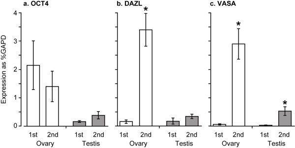
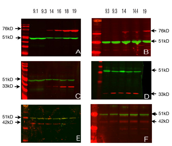
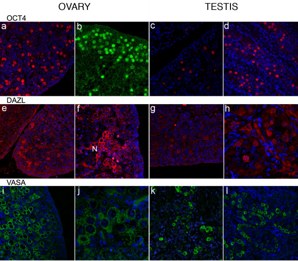
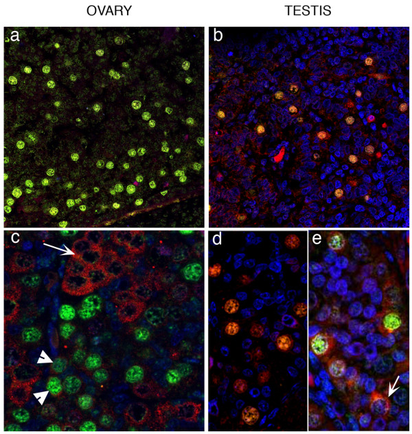
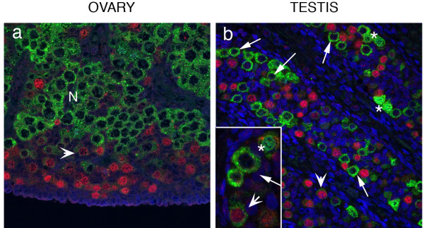
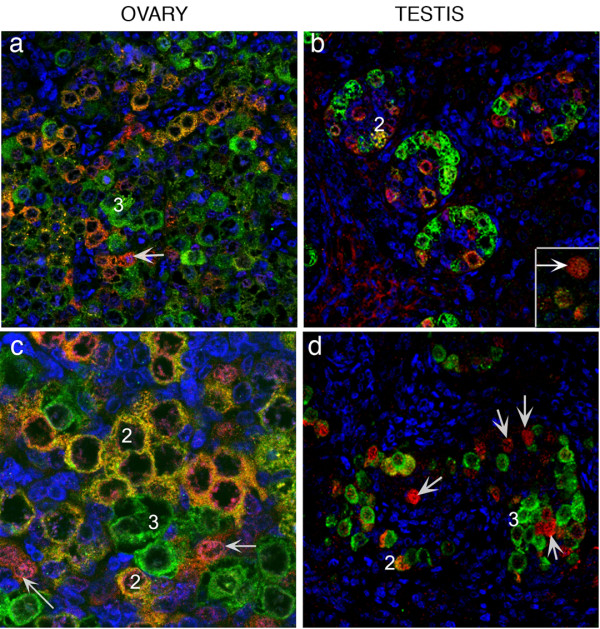
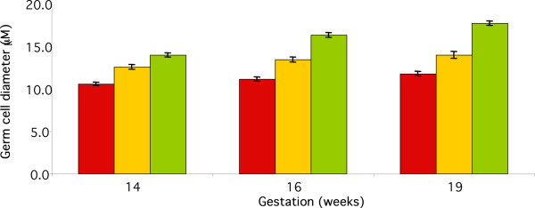
Results: OCT4 mRNA and protein were detected in extracts from both 1st and 2nd trimester ovaries and testes. In ovarian extracts a marked increase in expression of VASA and DAZL mRNA and protein occurred in the 2nd trimester. In testicular extracts VASA mRNA and protein were low/undetectable in 1st trimester and increased in the 2nd trimester whereas the total amount of DAZL did not seem to change. During the 1st trimester, germ cells were OCT4 positive but did not express VASA. These results are in contrast to the situation in mice where expression of Vasa is initiated in Oct4 positive primordial germ cells as they enter the gonadal ridge. In the 2nd trimester germ cells with intense cytoplasmic staining for VASA were present in both sexes; these cells were OCT4 negative. DAZL expression overlapped with both OCT4 and VASA and changed from the nuclear to the cytoplasmic compartment as cells became OCT4-negative. In males, OCT4-positive and VASA-positive subpopulations of germ cells coexisted within the same seminiferous cords but in the ovary there was a distinct spatial distribution of cells with OCT4 expressed by smaller, peripherally located, germ cells whereas DAZL and VASA were immunolocalised to larger (more mature) centrally located cells.

Conclusion: OCT4, DAZL and VASA are expressed by human fetal germ cells but their patterns of expression are temporally and spatially distinct. In the 1st trimester OCT4 was detected in most germ cells. In the 2nd trimester the onset of expression of VASA was associated with the formation of oocytes and spermatogonia both of which were OCT-4 negative. Relocation of DAZL from nucleus to cytoplasm paralleled the down regulation of OCT4 and the onset of expression of VASA. These data reveal similarities between the expression of key regulatory proteins within germ cells as they mature in male and female fetal human gonads suggesting that in the female these maturational changes are not determined by entry into meiosis.


Figures

Figure 1
Figure 1
Expression of mRNAs for OCT4, DAZL and VASA in 1st and 2nd trimester ovary and testis. Open columns, 1st trimester, shaded columns 2nd trimester. Concentrations of mRNA are all relative to that of GAPD in the same samples. * P < 0.001 vs 1st trimester. Mean ± sem, n = 5–18 per group.
Figure 2
Figure 2
Western analysis of 1st and 2nd trimester ovaries and testes. In both ovarian (A) and testicular (B) samples VASA (76 Kd) was not detectable in the 1st trimester samples but was present in those from the 2nd trimester. DAZL (33 Kd) was low/undetectable in 1st trimester ovaries (C) whereas it was detectable in ovarian samples from 2nd trimester and testicular samples from both 1st and 2nd trimester (D). OCT4 (42 Kd) was present in both ovaries (E) and testes (F) during both the 1st and 2nd trimester. The loading control in all cases was β-tubulin (51 Kd).
Figure 3
Figure 3
Immunoexpression of OCT4, DAZL and VASA. OCT4 positive germ cell nuclei were detectable in both the 1st and 2nd trimester ovaries (a, 62 d; b, 16 wk) and testes (c, 64 d; d, 16 wk). DAZL positive germ cells were rare in the 1st trimester (e, ovary 61 d; g, testis 64 d) but groups of cells ('nests', labelled N) with cytoplasmic staining were present in the 2nd trimester ovaries (f, 20 wk). During the 2nd trimester VASA protein was detected in the cytoplasm of female germ cells (i, 14 wk; j, 18 wk) throughout the ovary with the exception of the sub-epithelial layer. In the testes (k, 15 wk; l, 16 wk) VASA-positive germ cells were found in all cords.
Figure 4
Figure 4
Co-localisation of OCT4 and DAZL. In 1st trimester ovaries (a, 61 d) and testes (b, 64 d) OCT4 (green) and DAZL (red) were co-localised to germ cell nuclei. In ovaries from the 2nd trimester (c, 14 wk) DAZL protein was almost exclusively cytoplasmic and was largely localised to OCT4 negative groups of cells (arrow); a few OCT4 positive cells had a low level of DAZL immunoexpression in their cytoplasm (arrowheads). Testes, panel b, 64 d; panel d, 16 wk; panel e, 19 wk gestation. In 2nd trimester testes (d, 16 wk; e, 19 wk) DAZL was still expressed in the nuclei of some OCT4 positive germ cells but this pattern of expression was variable with DAZL protein present in the cytoplasm of OCT4 positive and OCT4 negative (arrow panel e) cells.
Figure 5
Figure 5
Co-localisation of OCT4 and VASA in 16 week ovary and testes. Panel a, ovary, cells with intense immunopositive staining for OCT4 are found at the periphery of the organ (red nuclei), VASA was detected in the cytoplasm and was most intense in cells located in nests (N) closer to the centre of the ovary. An intermediate population of cells with low intensity nuclear staining for OCT4 and low intensity staining for VASA (arrowheads) was also present. Panel b, testis, OCT4 positive and VASA positive germ cells were found within the same seminiferous cords; germ cells with intense nuclear OCT4 expression (red nuclei) were VASA negative and those with intense cytoplasmic expression of VASA (e.g. arrowed in inset panel b) were OCT 4 negative. Two other populations of male germ cells were identified, a population with low intensity immunoexpression of OCT4 which also had low intensity staining for VASA (arrowheads) and cells with nuclear VASA expression (asterisks) which were typically found in pairs.
Figure 6
Figure 6
Co-localisation of DAZL and VASA in 2nd trimester ovary and testes. In the 2nd trimester (ovary, a, 17 wk; c, 14 wk; testes b, 15 wk; d, 16 wk) three populations of germ cells were identified based on their patterns of expression of DAZL (red) and VASA (green). 1stly a rare population of cells which had nuclear DAZL (arrows), 2ndly cells which co-expressed both proteins (population 2) and thirdly the most prevalent group which were VASA positive/DAZL negative (population 3). Although few cells in population 2 were detected in testes these germ cells were found as distinct groups within the ovary (see panel c).
Figure 7
Figure 7
Ovarian germ cell diameter for cells expressing DAZL, VASA or both at 14, 16 and 19 weeks gestation. There were significant increases in diameter with gestation for each group (p = 0.001 for DAZL and VASA separately, p = 0.01 for cells expressing both). There were also significant differences (P < 0.01) between the 3 classifications of germ cell at each gestation. Mean ± sem, n = 20–120 per group.

References

    1. Brennan J, Capel B. One tissue, two fates: molecular genetic events that underlie testis versus ovary development. Nat Rev Genet. 2004;5:509–521. doi: 10.1038/nrg1381. - DOI - PubMed
    1. Saitou M, Barton SC, Surani MA. A molecular programme for the specification of germ cell fate in mice. Nature. 2002;418:293–300. doi: 10.1038/nature00927. - DOI - PubMed
    1. Wartenberg H. Differentiation and development of the testes. In: Burger H, de Kretser DM, editor. The Testis. 2nd. New York , Raven Press; 1981. pp. 39–81. (Comprehensive Endocrinology). Martini L.
    1. Rosner MH, Vigano MA, Ozato K, Timmons PM, Poirier F, Rigby PW, Staudt LMA. A POU-domain transcription factor in early stem cells and germ cells of the mammalian embryo. Nature. 1990;345:686–692. doi: 10.1038/345686a0. - DOI - PubMed
    1. Chambers I, Colby D, Robertson M, Nichols J, Lee S, Tweedie S, Smith A. Functional expression cloning of Nanog, a pluripotency sustaining factor in embryonic stem cells. Cell. 2003;113:643–655. doi: 10.1016/S0092-8674(03)00392-1. - DOI - PubMed

MeSH terms

LinkOut - more resources