

Differential effects of Th1, monocyte/macrophage and Th2 cytokine mixtures on early gene expression for glial and neural-related molecules in central nervous system mixed glial cell cultures: neurotrophins, growth factors and structural proteins
- PMID: 18088439
- PMCID: PMC2228280
- DOI: 10.1186/1742-2094-4-30
Differential effects of Th1, monocyte/macrophage and Th2 cytokine mixtures on early gene expression for glial and neural-related molecules in central nervous system mixed glial cell cultures: neurotrophins, growth factors and structural proteins
Abstract
Background: In multiple sclerosis, inflammatory cells are found in both active and chronic lesions, and it is increasingly clear that cytokines are involved directly and indirectly in both formation and inhibition of lesions. We propose that cytokine mixtures typical of Th1 or Th2 lymphocytes, or monocyte/macrophages each induce unique molecular changes in glial cells.
Methods: To examine changes in gene expression that might occur in glial cells exposed to the secreted products of immune cells, we have used gene array analysis to assess the early effects of different cytokine mixtures on mixed CNS glia in culture. We compared the effects of cytokines typical of Th1 and Th2 lymphocytes and monocyte/macrophages (M/M) on CNS glia after 6 hours of treatment.
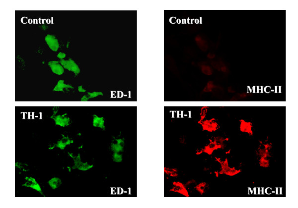
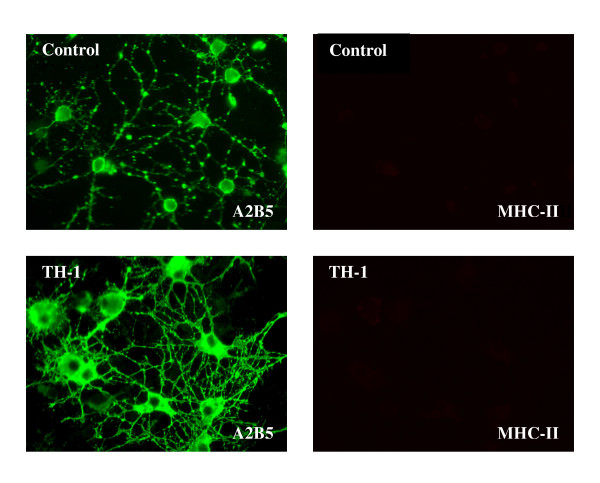
Results: In this paper we focus on changes with potential relevance for neuroprotection and axon/glial interactions. Each mixture of cytokines induced a unique pattern of changes in genes for neurotrophins, growth and maturation factors and related receptors; most notably an alternatively spliced form of trkC was markedly downregulated by Th1 and M/M cytokines, while Th2 cytokines upregulated BDNF. Genes for molecules of potential importance in axon/glial interactions, including cell adhesion molecules, connexins, and some molecules traditionally associated with neurons showed significant changes, while no genes for myelin-associated genes were regulated at this early time point. Unexpectedly, changes occurred in several genes for proteins initially associated with retina, cancer or bone development, and not previously reported in glial cells.
Conclusion: Each of the three cytokine mixtures induced specific changes in gene expression that could be altered by pharmacologic strategies to promote protection of the central nervous system.
Figures
Similar articles
-
Differential effects of Th1, monocyte/macrophage and Th2 cytokine mixtures on early gene expression for molecules associated with metabolism, signaling and regulation in central nervous system mixed glial cell cultures.J Neuroinflammation. 2009 Jan 21;6:4. doi: 10.1186/1742-2094-6-4. J Neuroinflammation. 2009. PMID: 19159481 Free PMC article.
-
Cytokines regulate neuronal gene expression: differential effects of Th1, Th2 and monocyte/macrophage cytokines.J Neuroimmunol. 2011 Sep 15;238(1-2):19-33. doi: 10.1016/j.jneuroim.2011.06.010. Epub 2011 Jul 30. J Neuroimmunol. 2011. PMID: 21803433
-
Differential effects of Th1, monocyte/macrophage and Th2 cytokine mixtures on early gene expression for immune-related molecules by central nervous system mixed glial cell cultures.Mult Scler. 2006 Apr;12(2):149-68. doi: 10.1191/135248506ms1251oa. Mult Scler. 2006. PMID: 16629418
-
TH1/TH2 cytokines in the central nervous system.Int J Neurosci. 2002 Jun;112(6):665-703. doi: 10.1080/00207450290025725. Int J Neurosci. 2002. PMID: 12325311 Review.
-
Cross-talk signals in the CNS: role of neurotrophic and hormonal factors, adhesion molecules and intercellular signaling agents in luteinizing hormone-releasing hormone (LHRH)-astroglial interactive network.Front Biosci. 1997 Mar 1;2:d88-125. doi: 10.2741/a177. Front Biosci. 1997. PMID: 9159216 Review.
Cited by
-
Serum and lymphocytic neurotrophins profiles in systemic lupus erythematosus: a case-control study.PLoS One. 2013 Nov 1;8(11):e79414. doi: 10.1371/journal.pone.0079414. eCollection 2013. PLoS One. 2013. PMID: 24223945 Free PMC article.
-
The interplay of inflammation and remyelination: rethinking MS treatment with a focus on oligodendrocyte progenitor cells.Mol Neurodegener. 2024 Jul 12;19(1):53. doi: 10.1186/s13024-024-00742-8. Mol Neurodegener. 2024. PMID: 38997755 Free PMC article. Review.
-
Pancreatic organogenesis mapped through space and time.Exp Mol Med. 2025 Feb;57(1):204-220. doi: 10.1038/s12276-024-01384-y. Epub 2025 Jan 8. Exp Mol Med. 2025. PMID: 39779976 Free PMC article.
-
Inflammatory mechanisms underlying cortical injury in progressive multiple sclerosis.Neuroimmunol Neuroinflamm. 2021;8:111-133. doi: 10.20517/2347-8659.2020.35. Epub 2021 Jun 20. Neuroimmunol Neuroinflamm. 2021. PMID: 40740587 Free PMC article.
-
Brain-derived neurotrophic factor serum levels and genotype: association with depression during interferon-α treatment.Neuropsychopharmacology. 2013 May;38(6):985-95. doi: 10.1038/npp.2012.263. Epub 2012 Dec 18. Neuropsychopharmacology. 2013. PMID: 23303061 Free PMC article.
References
-
- Brosnan CF, Cannella B, Battistini L, Raine CS. Cytokine localization in multiple sclerosis lesions: correlation with adhesion molecule expression and reactive nitrogen species. Neurology. 1995;45:S16–21. - PubMed
Publication types
MeSH terms
Substances
LinkOut - more resources
Full Text Sources
Molecular Biology Databases
