. 2008;187(3):199-210.
doi: 10.1159/000112640. Epub 2007 Dec 17.

Dynamic analysis of the expression of the TGFbeta/SMAD2 pathway and CCN2/CTGF during early steps of tooth development

Affiliations

Dynamic analysis of the expression of the TGFbeta/SMAD2 pathway and CCN2/CTGF during early steps of tooth development

Marcos S Pacheco et al. Cells Tissues Organs. 2008.

Abstract

Background/aims: CCN2 is present during tooth development. However, the relationship between CCN2 and the transforming growth factor beta (TGFbeta)/SMAD2/3 signaling cascade during early stages of tooth development is unclear. Here, we compare the expression of CCN2 and TGFbeta/SMAD2/3 components during tooth development, and analyze the functioning of TGFbeta/SMAD2/3 in wild-type (WT) and Ccn2 null (Ccn2-/-) mice.

Methods: Coronal sections of mice on embryonic day (E)11.5, E12.5, E13.5, E14.5 and E18.5 from WT and Ccn2-/- were immunoreacted to detect CCN2 and components of the TGFbeta signaling pathway and assayed for 5'-bromo-2'-deoxyuridine immunolabeling and proliferating cell nuclear antigen immunostaining.

Results: CCN2 and TGFbeta signaling components such as TGFbeta1, TGFbeta receptor II, SMADs2/3 and SMAD4 were expressed in inducer tissues during early stages of tooth development. Proliferation analysis in these areas showed that epithelial cells proliferate less than mesenchymal cells from E11.5 to E13.5, while at E14.5 they proliferate more than mesenchymal cells. We did not find a correlation between functioning of the TGFbeta1 cascade and CCN2 expression because Ccn2-/- mice showed neither a reduction in SMAD2 phosphorylation nor a difference in cell proliferation.

Conclusion: CCN2 and the TGFbeta/SMAD2/3 signaling pathway are active in signaling centers of tooth development where proliferation is dynamic, but these mechanisms may act independently.


Figures

Fig. 1
Fig. 1
CCN2 can be specifically detected in dental tissues by immunohistochemistry. a WT molar tooth at E13.5 reacted with anti-CCN2 antibody. CCN2 staining is detected in condensed mesenchyme and in Meckel’s cartilage. b CCN2−/− immunostained with anti-CCN2 antibody showing no specific expression. c E18.5 reacted with anti-CCN2 antibody. CCN2 staining is detected in dental lamina (asterisk) and inner epithelium (arrowhead). d Negative reaction where anti-CCN2 polyclonal antibody was omitted showing no specific expression. e Inset showing Ccn2−/− at E18.5 when immunostained with anti-CCN2 antibody showing no specific expression. B = Buccal side; L = lingual side. Scale bars: 50 μm (a–d) and 100 μm (e).
Fig. 2
Fig. 2
CCN2 is expressed in signaling centers during early stages of tooth development. a At E11.5 immuno-staining is detected in dental lamina and in a few cells in underlying mesenchyme. c At E13.5 (bud stage) staining of CCN2 is detected in condensed mesenchyme around the epithelial bud. Non-dental epithelial cells also express CCN2. Non-condensed mesenchyme appeared almost completely unlabeled. e At E14.5 (cap stage) CCN2 is detected in dental lamina, inner and outer epithelium, enamel knot and dental sac. CCN2 can also be detected in some cells in non-condensed mesenchyme. b, d, f Drawing of the staining shown on the left panels: dots in red indicate cells expressing CCN2. On the left side embryonic stages and section plan is shown. B = Buccal side; CM = condensed mesenchyme; DL = dental lamina; EK = enamel knot; IE = inner epithelium; L = lingual side; ME = mesenchyme; NCM = non-condensed mesenchyme; OE = outer epithelium; SR = stellate reticulum; TB = tooth bud. Scale bars = 50 μm.
Fig. 3
Fig. 3
TGFβ1 and TGFβRII are similarly expressed in signaling centers during early steps of tooth development. At E11.5 TGFβ1 (a) and TGFβRII (d) expression are weakly detected in dental lamina and in a few cells in underlying mesenchyme. At E13.5 TGFβ1 (b) and TGFβRII (e) are expressed in condensed mesenchyme and in cells of the epithelial bud closer to the condensed mesenchyme. At E14.5, TGFβ1 (c) and TGFβRII (f) are highly expressed in inner epithelium, outer epithelium, enamel knot, and are also detected in a few cells in the condensed mesenchyme. B = Buccal side; L = lingual side. Scale bars = 50 μm.
Fig. 4
Fig. 4
SMAD2/3 and SMAD4 are similarly expressed in signaling centers during early stages of tooth development. a At E11.5 SMAD2/3 is strongly stained in dental lamina and a few cells can be detected amongst the mesenchymal cells next to the dental lamina. d SMAD4 expression at E11.5 is mainly detected in dental lamina, however it is weaker than SMAD2/3. SMAD4 is also detected in mesenchymal cells. b At E13.5, SMAD2/3 and SMAD4 (e) expression occurs in condensed mesenchyme and in epithelial bud cells. At E14.5, SMAD2/3 (c) and SMAD4 (f) expression are similarly expressed in outer epithelium, inner epithelium and enamel knot. Weak expression of SMAD2/3 and SMAD4 can be detected in non-condensed mesenchyme and cells of condensed mesenchyme are also stained. B = Buccal side; L = lingual side. Scale bar = 50 μm.
Fig. 5
Fig. 5
BrdU and PCNA analysis of cell proliferation during early stages of tooth development. a, b At E11.5 BrdU-positive cells are present in underlying mesenchyme and in a few cells in dental lamina. At this stage, mesenchymal cells show higher proliferation than dental lamina. c Likewise, PCNA staining at E11.5 shows proliferation in dental lamina, and in cells in underlying mesenchyme. d, e At E12.5 expression of BrdU-positive cells is more prominent in condensed mesenchyme than in invaginating epithelium. f PCNA staining is intensely detected in condensed mesenchyme and can be detected at a lower intensity in invaginating epithelium. g, h At 13.5 BrdU-positive cells are detected in both epithelial bud and condensed mesenchyme. i PCNA staining confirms the same proliferation profile as the observed in BrdU labeling. Notice that proliferation is not highly detected in non-condensed mesenchyme. j, k At E14.5, BrdU-positive cells are detected in inner and outer epithelium. l Similarly, PCNA staining is also found in inner and outer epithelium, but not in enamel knot cells (arrow). B = Buccal side; DAPI = 4′-6-diamidino-2-phenylindole; L = lingual side. Scale bar = 50 μm.
Fig. 6
Fig. 6
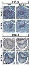
SMAD2 phosphorylation and proliferation is not affected in Ccn2 −/−. At E13.5, SMAD2P in both WT (a) and Ccn2−/− (b) is detected in invaginating epithelial cells and in condensed and non-condensed mesenchyme cells. PCNA staining in WT (c) and Ccn2−/− (d) is mainly detected in epithelium and in non-condensed mesenchyme but a few cells can be detected in condensed mesenchyme. At E18.5 SMAD2P is detected in stellate reticulum, inner epithelium and mesenchyme cells in WT (e) and Ccn2−/− (f). PCNA staining is mainly detected in stellate reticulum, but is less abundant in mesenchymal cells in both WT (g) and Ccn2−/− (h). Notice that no significant difference in SMAD2P and PCNA staining is found when WT is compared with Ccn2−/−. B = Buccal side; L = lingual side. Scale bar = 50 μm.

Similar articles

Cited by

References

    1. Abdollah S, Macias-Silva M, Tsukazaki T, Hayashi H, Attisano L, Wrana JL. TGFβRI phosphorylation Smad2 on Ser465 and Ser467 is required for Smad2-Smad4 complex formation and signaling. J Biol Chem. 1997;272:27678–27685. - PubMed
    1. Abreu JG, Coffinier C, Larraín J, Oelge-schlager M, De Robertis EM. Chordin-like CR and the evolutionarily conserved extracellular signaling system. Gene. 2002a;287:39–47. - PubMed
    1. Abreu JG, Ketpura NI, Reversade B, De Robertis EM. Connective-tissue growth factor (CTGF) modulates cell signaling by BMP and TGF-β. Nat Cell Biol. 2002b;4:599–604. - PMC - PubMed
    1. Bork P. The modular architecture of a new family of growth regulators related to connective tissue growth factor. FEBS Lett. 1993;327:125–130. - PubMed
    1. Brunner A, Chinn J, Neubauer M, Purchio AF. Identification of a gene family regulated by transforming growth factor-β. DNA Cell Biol. 1991;10:293–300. - PubMed

Publication types

MeSH terms