. 2007 Dec;66(12):1136-46.
doi: 10.1097/nen.0b013e31815c5efb.

Clinicopathologic correlations in a large Alzheimer disease center autopsy cohort: neuritic plaques and neurofibrillary tangles "do count" when staging disease severity

Affiliations

Clinicopathologic correlations in a large Alzheimer disease center autopsy cohort: neuritic plaques and neurofibrillary tangles "do count" when staging disease severity

Peter T Nelson et al. J Neuropathol Exp Neurol. 2007 Dec.

Abstract

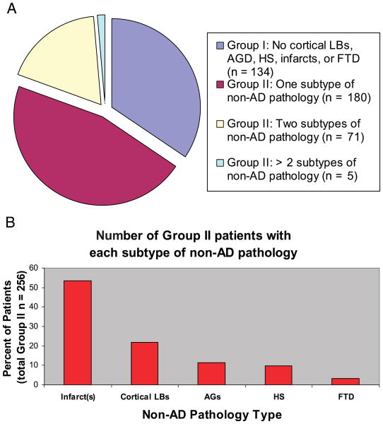
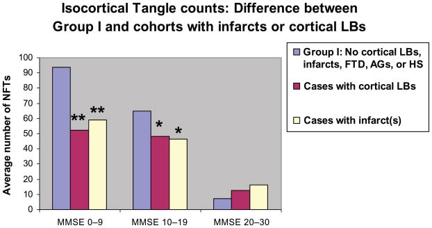
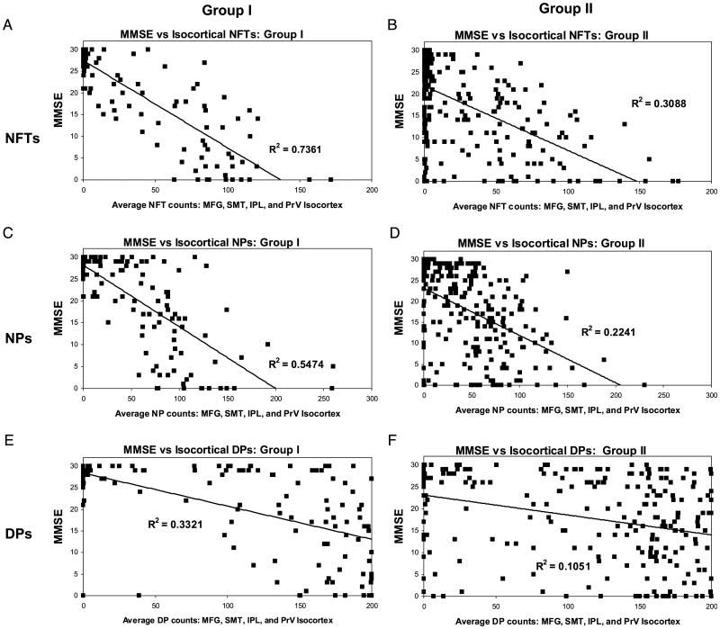
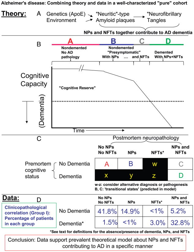
There is uncertainty regarding the association of cognitive decline in Alzheimer disease (AD) with classic histopathologic features- neurofibrillary tangles (NFTs) and "neuritic" amyloid plaques (NPs). This uncertainty fuels doubts about the diagnostic importance of NFTs and NPs and leads to confusion regarding hypotheses of AD pathogenesis. Three hundred ninety subjects who underwent longitudinal premortem clinical workup and postmortem quantitative neuropathologic assessment served as the group to address this issue. Subjects with concomitant brain disease(s) were analyzed independently to more accurately assess the contribution of distinct pathologies to cognitive decline. More than 60% of patients of all age groups had important non-AD brain pathologies. However, subjects without superimposed brain diseases showed strong correlations between AD-type pathology counts (NFTs > NPs) and premortem Mini-Mental State Examination scores. The observed correlation was stronger in isocortex than in allocortex and was maintained across age groups including patients older than 90 years. A theoretical model is proposed in which our results are interpreted to support the "amyloid cascade hypothesis" of AD pathogenesis. Our data show that there are many important contributory causes to cognitive decline in older persons. However, NFTs and NPs should not be dismissed as irrelevant in AD based on clinicopathologic correlation.


Figures

FIGURE 1
FIGURE 1
Histopathologic hallmarks of Alzheimer disease (AD). A neurofibrillary tangle (diagonal arrows) and neuritic plaque (oval-shaped structure on right) are shown in a photomicrograph from human AD brain section stained with the silver-impregnation Bielschowsky technique. Neurofibrillary tangles are composed of insoluble and protease-resistant fibrils, and develop intracellularly. Neuritic plaques are extracellular fibrillary amyloid deposits, surrounded by swollen, degenerating, silver-impregnated neurites. Scale bar = 50 μm.
FIGURE 2
FIGURE 2
(A) Almost two-thirds of aged human brains contain non-Alzheimer disease (AD)-type neuropathology. (B) The subtypes of non-AD pathology are shown in order of prevalence. In the cohort seen at the University of Kentucky Alzheimer's Disease Center, cerebral infarcts are the most prevalent non-AD pathology, followed by cortical Lewy bodies.
FIGURE 3
FIGURE 3
Patients with impaired cognition and findings of Alzheimer disease (AD) only have more neurofibrillary tangles (combined counts from superior and middle temporal gyri, middle frontal gyrus, inferior parietal lobule, and occipital lobe including the primary visual area) than cohorts with similar levels of cognitive function and concurrent non-AD pathologic findings (Group II). Patients are grouped according to MMSE scores. Blue bars represent Group I no Lewy bodies (LBs), argyrophilic grains (AGs), infarct(s), hippocampal sclerosis (HS), or frontotemporal dementia (FTD). The red bars and yellow bars refer to all Group II cases with cortical LBs or infarct(s), respectively. Results of 2-tailed, 2-sample Student t-test: *, p < 0.05; **, p < 0.001.
FIGURE 4
FIGURE 4
Correlating histopathologic lesions of Alzheimer disease (AD) with premortem cognitive decline as quantified with the Mini-Mental State Examination (MMSE). (A, B) Results for neurofibrillary tangles (NFTs), (C, D) results for neuritic amyloid plaques (NPs), and (E, F) results for diffuse plaques (DPs) in Group I (A, C, E) and Group II (with Lewy bodies, argyrophilic grains, infarct[s], hippocampal sclerosis, and/or frontotemporal dementia) (B, D, F). Note that the correlation coefficients are consistently higher for Group I versus Group II, and for both groups the correlation coefficients are NFTs > NPs > DPs. There are a few Group I patients with dementia who lack NFTs and NPs. SMT, superior and middle temporal gyri; MFG, middle frontal gyrus; IPL, inferior parietal lobule; PrV, primary visual cortex.
FIGURE 5
FIGURE 5
Clinical and pathologic data are assessed in the context of prevalent hypotheses about the pathogenesis of Alzheimer disease (AD). (A) The amyloid cascade hypothesis is that genetic and environmental influences contribute to the formation of neuritic amyloid plaques (NPs), which in turn potentiate neurofibrillary tangle (NFT) formation, and together, NFTs and NPs contribute to the synapse elimination and cell death seen in AD. (B) The chart relates the clinical features of AD to the pathology. As NPs and NFTs accumulate and cognition deteriorates there is a time during which no overt dementia is detectable. (C) A simple chart to evaluate whether the hypotheses about AD pathogenesis are borne out by clinicopathologic correlation. (D) It would be counter to the hypotheses in A and B, if the table positions in C designated w, x, y, and z were highly represented in the population. (E) Empirical data from Group I patients are highly compatible with the hypothesis shown in A and B, because the vast majority of patients either have no dementia with no pathology (∼42%), have dementia with NPs and NFTs (∼33%), or are in some transitional condition (∼20%).

Similar articles

Cited by

References

    1. Alzheimer A. Uber egenartige Krankheitsfalle des spateren Alters. Z Ges Neurol. 1911;4:356–85.
    1. Lee HG, Zhu X, Castellani RJ, et al. Amyloid-β in Alzheimer disease: The null versus the alternate hypotheses. J Pharmacol Exp Ther. 2007;321:823–29. - PubMed
    1. Wirths O, Multhaup G, Bayer TA. A modified β-amyloid hypothesis: Intraneuronal accumulation of the β-amyloid peptide—the first step of a fatal cascade. J Neurochem. 2004;91:513–20. - PubMed
    1. Marchesi VT. An alternative interpretation of the amyloid Aβ hypothesis with regard to the pathogenesis of Alzheimer's disease. Proc Natl Acad Sci USA. 2005;102:9093–98. - PMC - PubMed
    1. Lesne S, Koh MT, Kotilinek L, et al. A specific amyloid-β protein assembly in the brain impairs memory. Nature. 2006;440:352–57. - PubMed

Publication types

MeSH terms

Substances