. 2007:3:152.
doi: 10.1038/msb4100200. Epub 2007 Dec 18.

A map of human cancer signaling

Affiliations

A map of human cancer signaling

Qinghua Cui et al. Mol Syst Biol. 2007.

Abstract

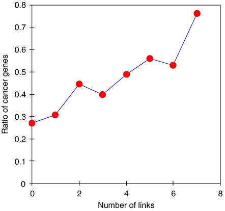
We conducted a comprehensive analysis of a manually curated human signaling network containing 1634 nodes and 5089 signaling regulatory relations by integrating cancer-associated genetically and epigenetically altered genes. We find that cancer mutated genes are enriched in positive signaling regulatory loops, whereas the cancer-associated methylated genes are enriched in negative signaling regulatory loops. We further characterized an overall picture of the cancer-signaling architectural and functional organization. From the network, we extracted an oncogene-signaling map, which contains 326 nodes, 892 links and the interconnections of mutated and methylated genes. The map can be decomposed into 12 topological regions or oncogene-signaling blocks, including a few 'oncogene-signaling-dependent blocks' in which frequently used oncogene-signaling events are enriched. One such block, in which the genes are highly mutated and methylated, appears in most tumors and thus plays a central role in cancer signaling. Functional collaborations between two oncogene-signaling-dependent blocks occur in most tumors, although breast and lung tumors exhibit more complex collaborative patterns between multiple blocks than other cancer types. Benchmarking two data sets derived from systematic screening of mutations in tumors further reinforced our findings that, although the mutations are tremendously diverse and complex at the gene level, clear patterns of oncogene-signaling collaborations emerge recurrently at the network level. Finally, the mutated genes in the network could be used to discover novel cancer-associated genes and biomarkers.


Figures

Figure 1
Figure 1
Illustration of the sources of cancer mutated network genes and oncogenic signal transduction events. (A) Most of the cancer mutated network genes were discovered by large-scale sequencing of tumor samples, whereas a small fraction of them was found in literature. (B) Oncogenic signal transduction events and oncogene-signaling-dependent events. (a) Signaling divergent unit. The line in red represents an oncogenic signal transduction event. (b) Signaling convergent unit. The line in red represents an oncogene-signaling-dependent event. In this case, both genes have high mutation frequency (⩾0.02), suggesting that the signaling event between the two genes is frequently used in tumorigenesis. Nodes in red represent mutated genes, whereas numbers represent mutation frequencies. Signs + and − represent activating and inhibitory links, respectively.
Figure 2
Figure 2
Enrichment of mutated and methylated genes in network motifs. (A) Relations between the fractions of positive links in all 3-node-size network motifs and the fractions of mutated genes in these motifs. (B) Relations between the fractions of positive links in all 3-node-size network motifs and the fractions of methylated genes in these motifs. All network motifs were classified into subgroups based on the number of nodes that are either mutated genes or methylated genes, respectively. The ratio of positive links to total positive and negative links in each subgroup was plotted. The horizontal lines indicate the ratio of positive links to the total positive and negative links in all network motifs.
Figure 3
Figure 3
Human oncogene-signaling map. The human cancer-signaling map was extracted from the human signaling network, which was mapped with cancer mutated and methylated genes. The map shows three ‘oncogenic-dependent regions' (background in light gray), in which genes of the two regions are also heavily methylated. Nodes represent genes, whereas the links with and without arrows represent signal and physical relations, respectively. Nodes in red, purple, brown, cyan, blue and green represent the genes that are highly mutated but not methylated, both highly mutated and methylated, poorly mutated but not methylated, both poorly mutated and methylated, methylated but not mutated, and neither mutated nor methylated, respectively.
Figure 4
Figure 4
Heatmaps of the gene mutation distributions in oncogene-signaling blocks. Twelve topological regions or oncogene-signaling blocks have been identified based on the gene connectivity of the human oncogene-signaling map. A heatmap was generated from a matrix, which was built by querying the oncogene signaling blocks using tumor samples, in which each sample has at least two mutated genes. If a gene of a particular signaling block (b) gets mutated in a tumor sample (s), we set Ms,b to 1, otherwise we set Ms,b to 0. (A) A heatmap generated using the gene mutation data of the 592 tumor samples. (B) A heatmap generated using the gene mutation data of the NCI-60 cancer cell lines. (C, D) Heatmaps generated using the output from the genome-wide sequencing of breast and colon tumor samples, respectively. Rows represent samples, whereas columns represent oncogene-signaling blocks. Samples were organized according to the cancer types they belong to. Cancer types that have relatively more samples were marked on the heatmap: (a) breast, (b) central nervous system, (c) blood, (d) lung, (e) pancreas and (f) skin tumors. Blocks with gene mutations are marked in yellow; however, when one sample contains statistically significant co-occurring mutated gene pairs (see Supplementary Table 7), the blocks are marked in red.
Figure 5
Figure 5
Heatmaps of the gene mutation distributions in oncogene-signaling blocks for six representative cancer types. Heatmaps for (A) blood, (B) breast, (C) central nervous system, (D) lung, (E) pancreas and (F) skin tumors were built using tumor samples of these cancer types, respectively. Rows represent samples, whereas columns represent oncogene-signaling blocks. Blocks with gene mutations are marked in yellow; however, when one sample contains statistically significant co-occurring mutated gene pairs (see Supplementary Table 7), the blocks are marked in red.
Figure 6
Figure 6
Correlation between the link number of a gene to the mutated genes and cancer-associated genes. We first classified the network genes (without mutations) into groups based on the number of the cancer mutated genes a gene links to. We then calculated the ratio of the cancer-associated genes to total genes for each group. The correlation between the ratio and the groups was plotted.

References

    1. Awan A, Bari H, Yan F, Mokin S, Yang S, Chowdhury Q, Yu Z, Purisima EO, Wang E (2007) Regulatory network motifs and hotspots of cancer genes in a mammalian cellular signaling network. IET Syst Biol 1: 292–297 - PubMed
    1. Babu MM, Luscombe NM, Aravind L, Gerstein M, Teichmann SA (2004) Structure and evolution of transcriptional regulatory networks. Curr Opin Struct Biol 14: 283–291 - PubMed
    1. Chang HY, Nuyten DS, Sneddon JB, Hastie T, Tibshirani R, Sorlie T, Dai H, He YD, van't Veer LJ, Bartelink H, van de RM, Brown PO, van d V (2005) Robustness, scalability, and integration of a wound-response gene expression signature in predicting breast cancer survival. Proc Natl Acad Sci USA 102: 3738–3743 - PMC - PubMed
    1. Chanock SJ, Thomas G (2007) The devil is in the DNA. Nat Genet 39: 283–284 - PubMed
    1. Chng WJ (2007) Limits to the human cancer genome project? Science 315: 762–765 - PubMed

Publication types