. 2008;153(3):445-53.
doi: 10.1007/s00705-007-0007-4. Epub 2007 Dec 19.

Pathogenesis of bovine spongiform encephalopathy in sheep

Affiliations

Pathogenesis of bovine spongiform encephalopathy in sheep

L J M van Keulen et al. Arch Virol. 2008.

Abstract

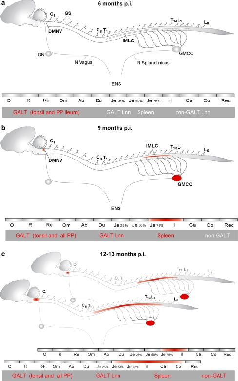
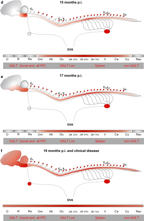
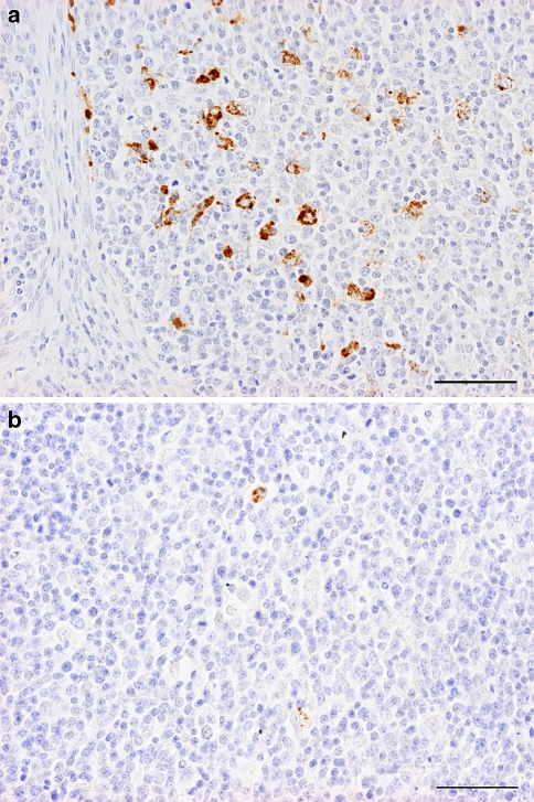
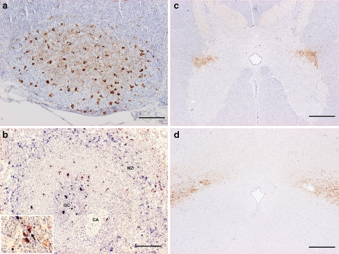
The pathogenesis of bovine spongiform encephalopathy (BSE) in sheep was studied by immunohistochemical detection of scrapie-associated prion protein (PrP(Sc)) in the gastrointestinal, lymphoid and neural tissues following oral inoculation with BSE brain homogenate. First accumulation of PrP(Sc) was detected after 6 months in the tonsil and the ileal Peyer's patches. At 9 months postinfection, PrP(Sc) accumulation involved all gut-associated lymphoid tissues and lymph nodes as well as the spleen. At this time point, PrP(Sc) accumulation in the peripheral neural tissues was first seen in the enteric nervous system of the caudal jejunum and ileum and in the coeliac-mesenteric ganglion. In the central nervous system, PrP(Sc) was first detected in the dorsal motor nucleus of the nervus Vagus in the medulla oblongata and in the intermediolateral column in the spinal cord segments T7-L1. At subsequent time points, PrP(Sc) was seen to spread within the lymphoid system to also involve all non-gut-associated lymphoid tissues. In the enteric nervous system, further spread of PrP(Sc) involved the neural plexi along the entire gastrointestinal tract and in the CNS the complete neuraxis. These findings indicate a spread of the BSE agent in sheep from the enteric nervous system through parasympathetic and sympathetic nerves to the medulla oblongata and the spinal cord.


Figures

Fig. 1
Fig. 1
Schematic representation of PrPSc accumulation (in red) in the lymphoid tissues, ENS and CNS of ARQ/ARQ sheep orally infected with BSE after a 6 months b 9 months c 12–13 months d 15 months e 17 months and f 19 months and after the onset of clinical disease. Abbreviations: DMNV dorsal motor nucleus of the vagus; IMLC intermediolateral column; C, T and L cervical, thoracic and lumbal segments of the spinal cord; GN ganglion nodosus; GMCC ganglion mesentericum cranialis/coeliacum; GS ganglia spinalis; O oesophagus; R rumen; Re reticulum; Om omasum; Ab abomasum; Du duodenum; Je jejunum; Il ileum; Ca caecum; Co colon; Rec rectum; GALT gut-associated lymphoid tissues; GALT Lnn lymph nodes draining from GALT tissues. Only the efferent motor fibers of the autonomic nervous system are shown
Fig. 1
Fig. 1
Schematic representation of PrPSc accumulation (in red) in the lymphoid tissues, ENS and CNS of ARQ/ARQ sheep orally infected with BSE after a 6 months b 9 months c 12–13 months d 15 months e 17 months and f 19 months and after the onset of clinical disease. Abbreviations: DMNV dorsal motor nucleus of the vagus; IMLC intermediolateral column; C, T and L cervical, thoracic and lumbal segments of the spinal cord; GN ganglion nodosus; GMCC ganglion mesentericum cranialis/coeliacum; GS ganglia spinalis; O oesophagus; R rumen; Re reticulum; Om omasum; Ab abomasum; Du duodenum; Je jejunum; Il ileum; Ca caecum; Co colon; Rec rectum; GALT gut-associated lymphoid tissues; GALT Lnn lymph nodes draining from GALT tissues. Only the efferent motor fibers of the autonomic nervous system are shown
Fig. 2
Fig. 2
BSE-infected sheep 6 months after oral inoculation. PrPSc immunostaining of a palatine tonsil and b Peyer’s patches of the ileum. PrPSc accumulations are present in tingible body macrophages of the lymphoid follicles. Bar 50 μm
Fig. 3
Fig. 3
BSE-infected sheep 9 months after oral inoculation. PrPSc immunostaining in a the Peyer’s patches of the ileum b the myenteric plexus of the ileum c IMLC of the spinal cord segment T10 and d DMNV at the obex. Note the sparse neuronal cell labelling in the IMLC (inlay in c) and the ventral part of the DMNV (inlay in d). aBar 50 μm; bbar 20 μm; c and d bar 500 μm
Fig. 4
Fig. 4
BSE-infected sheep 12 months after oral inoculation. PrPSc immunostaining in a the Peyer’s patches of the ileum b marginal zone in the spleen c IMLC of the spinal cord segment T5 and d DMNV at the obex . Note the colocalization of PrPSc (blue) and marginal zone macrophages (red) in the inlay in b (arrow). CA central arteriole; GC germinal center; MZ marginal zone. aBar 100 μm; bbar 100 μm; cbar 500 μm; dbar 500 μm

References

    1. Bellworthy SJ, Dexter G, Stack M, Chaplin M, Hawkins SA, Simmons MM, Jeffrey M, Martin S, Gonzalez L, Hill P (2005) Natural transmission of BSE between sheep within an experimental flock. Vet Rec 157:206 - PubMed
    1. Bellworthy SJ, Hawkins SA, Green RB, Blamire I, Dexter G, Dexter I, Lockey R, Jeffrey M, Ryder S, Berthelin Baker C, Simmons MM (2005) Tissue distribution of bovine spongiform encephalopathy infectivity in Romney sheep up to the onset of clinical disease after oral challenge. Vet Rec 156:197–202 - PubMed
    1. Bruce ME, Chree A, McConnell I, Foster JD, Pearson GR, Fraser H (1994) Transmission of bovine spongiform encephalopathy and scrapie to mice:strain variation and the species barrier. Phil Trans R Soc Lond B 343:405–411 - DOI - PubMed
    1. Bruce ME, Will RG, Ironside JWW, McConnell I, Drummond D, Suttie A, McCardle L, Chree A, Hope J, Birkett CR, Cousens SN, Fraser H, Bostock CJ (1997) Transmissions to mice indicate that ‘new variant’ CJD is caused by the BSE agent. Nature 389:498–501 - DOI - PubMed
    1. Buschmann A, Luhken G, Schultz J, Erhardt G, Groschup MH (2004) Neuronal accumulation of abnormal prion protein in sheep carrying a scrapie-resistant genotype (PrPARR/ARR). J Gen Virol 85:2727–2733 - DOI - PubMed

Publication types

MeSH terms