. 2007 Dec;26(12):1657-69.
doi: 10.1109/tmi.2007.901432.

Surface-constrained volumetric brain registration using harmonic mappings

Affiliations

Surface-constrained volumetric brain registration using harmonic mappings

Anand A Joshi et al. IEEE Trans Med Imaging. 2007 Dec.

Abstract

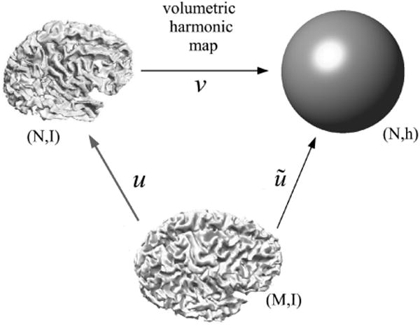
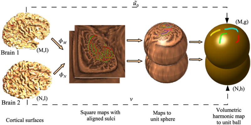
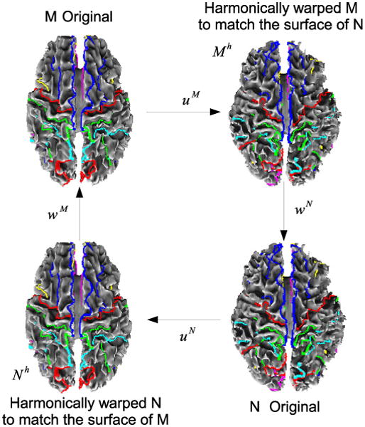
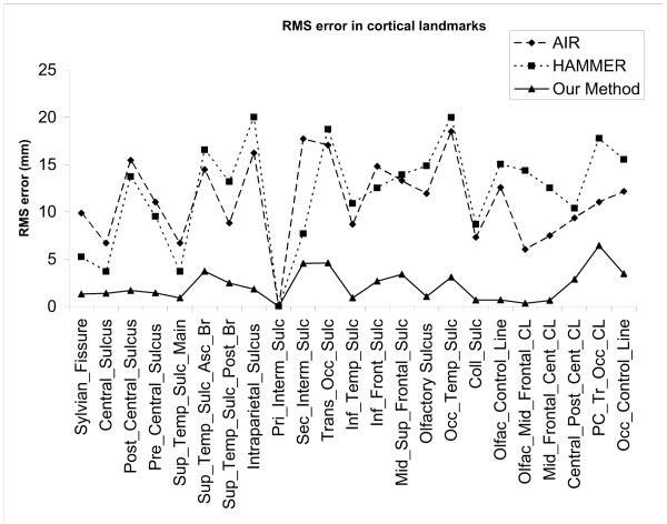
In order to compare anatomical and functional brain imaging data across subjects, the images must first be registered to a common coordinate system in which anatomical features are aligned. Intensity-based volume registration methods can align subcortical structures well, but the variability in sulcal folding patterns typically results in misalignment of the cortical surface. Conversely, surface-based registration using sulcal features can produce excellent cortical alignment but the mapping between brains is restricted to the cortical surface. Here we describe a method for volumetric registration that also produces an accurate one-to-one point correspondence between cortical surfaces. This is achieved by first parameterizing and aligning the cortical surfaces using sulcal landmarks. We then use a constrained harmonic mapping to extend this surface correspondence to the entire cortical volume. Finally, this mapping is refined using an intensity-based warp. We demonstrate the utility of the method by applying it to T1-weighted magnetic resonance images (MRIs). We evaluate the performance of our proposed method relative to existing methods that use only intensity information; for this comparison we compute the intersubject alignment of expert-labeled subcortical structures after registration.


Figures

Fig. 1
Fig. 1
Cortical surface alignment after using air software for intensity based volumetric alignment with a 168 parameter 5th order polynomial. Note that although the overall morphology is similar between the brains, the two cortical surfaces do not align well.
Fig. 2
Fig. 2
(a),(b) Two cortical surfaces with labeled sulci as colored curves; (c),(d) flat maps of a single hemisphere for each brains without the sulcal alignment constraint; (e),(f) flat maps with sulcal alignment; (g),(h) overlay of sulcal curves on the flat maps, without and with sulcal alignment.
Fig. 3
Fig. 3
Result of mapping of sulcal landmarks from 5 subjects to a single brain using the linear elastic mapping described here (left) without and (right) with the sulcal alignment constraint.
Fig. 4
Fig. 4
Illustration of our general framework for surface-constrained volume registration. We first compute the map v from brain manifold (N, I) to the unit ball to form manifold (N, h). We then compute a map ũ from brain (M, I) to (N, h). The final harmonic map from (M, I) to (N, I) is then given by u = v−1ũ.
Fig. 5
Fig. 5
Initialization for harmonic mapping from M to N. First we generate flat square maps of the two brains, one for each hemisphere, with pre-aligned sulci. The squares corresponding to each hemispheres are mapped to a disk and the disks are projected onto the unit sphere. We then generate a volumetric maps from each of the brains to the unit ball. Since all these maps are bijective, the resulting map results in a bijective point correspondence between the two brains. However, this correspondence is not optimal with respect to the harmonic energy of maps from the first brain to the second, but is used as an initialization for minimization of (20).
Fig. 6
Fig. 6
Illustration of the deformation induced with respect to the Euclidean coordinates by mapping to the unit ball. Shown are iso-surfaces corresponding to the Euclidean coordinates for different radii in the unit ball. Distortions become increasingly pronounced towards the outer edge of the sphere where the entire convoluted cortical surface is mapped to the surface of the ball.
Fig. 7
Fig. 7
Schematic of the intensity alignment procedure. Once harmonic maps uM and uN are computed, we refine these with intensity driven warps wM and wN while imposing constraints so that the final deformations are inverse consistent.
Fig. 8
Fig. 8
Illustration of the effects of the two stages of volumetric matching is shown by applying the deformations to a regular mesh representing one slice. Since the deformation is in 3d, the third in-paper value is represented by color. (a) Regular mesh representing one slice in the subject; (b) the regular mesh warped by the harmonic mapping which matches the subject cortical surface to the template cortical surface. Note that deformation is largest near the surface since the harmonic map is constrained only by the cortical surface; (c) Regular mesh representing one slice in the harmonically warped subject; (d) the intensity-based refinement now refines the deformation of the template to improve the match between subcortical structures. In this case the deformation is constrained to zero at the boundary and are confined to the interior of the volume.
Fig. 9
Fig. 9
Examples of surface constrained volumetric registration. (a) Original subject volume, original template, registration of subject to template using surface constrained harmonic mapping, intensity-based refinement of the harmonic map of subject to template is shown. Note that the surface of the warped subject matches to the surface of the template. (b) Anatomical labels of the subject and the template followed by labels of the subject warped by surface constrained harmonic mapping and intensity-based refinement of the harmonic map are shown.
Fig. 10
Fig. 10
RMS errors in alignment of different sulci using AIR (5th order), HAMMER and our surface constrained mapping.

References

    1. Christensen GE, Joshi SC, Miller MI. Volumetric transformation of brain anatomy. IEEE TMI. 1997 Dec;16(6) - PubMed
    1. Thompson PM, Toga AW. A surface-based technique for warping 3-dimensional brain. IEEE Transactions on Medical Imaging. 1996;15(4):1–16. - PubMed
    1. Talairach J, Tournoux P. Co-planar Stereotaxic Atlas of the Human Brain: 3-Dimensional Proportional System - an Approach to Cerebral Imaging. NY: Thieme Medical Publishers, New York; 1988.
    1. Ashburner J, Friston K. Spatial normalization. In: Toga A, editor. Brain Warping. Academic Press; 1999. pp. 27–44.
    1. Woods RP, Grafton ST, Holmes CJ, Cherry SR, Mazziotta JC. Automated image registration: I. General methods and intrasubject, intramodality validation. Journal of Computer Assisted Tomography. 1998;22:139–152. - PubMed

Publication types