

Phosphorylation of paxillin LD4 destabilizes helix formation and inhibits binding to focal adhesion kinase
- PMID: 18092823
- PMCID: PMC4054611
- DOI: 10.1021/bi702103n
Phosphorylation of paxillin LD4 destabilizes helix formation and inhibits binding to focal adhesion kinase
Abstract
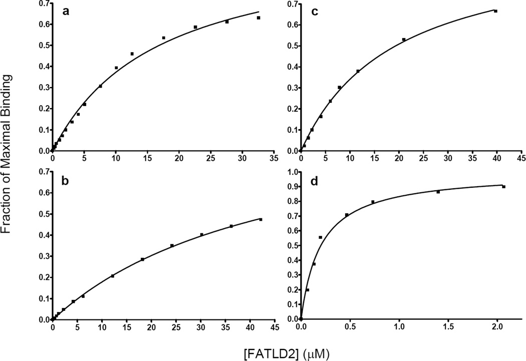
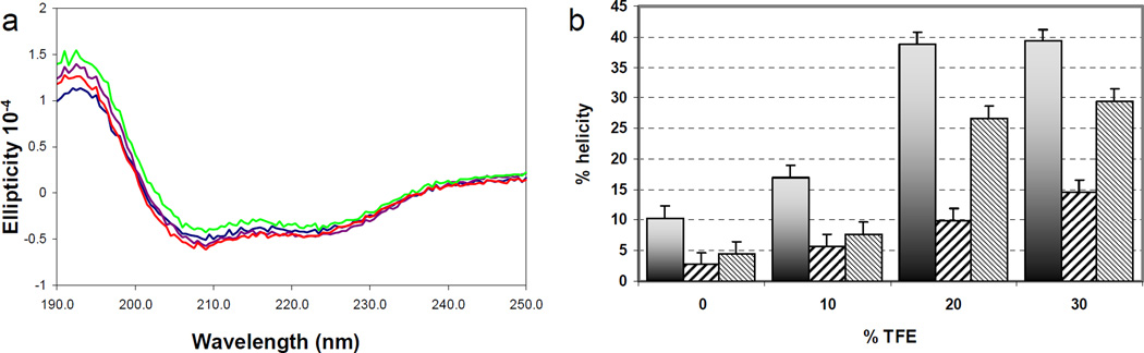
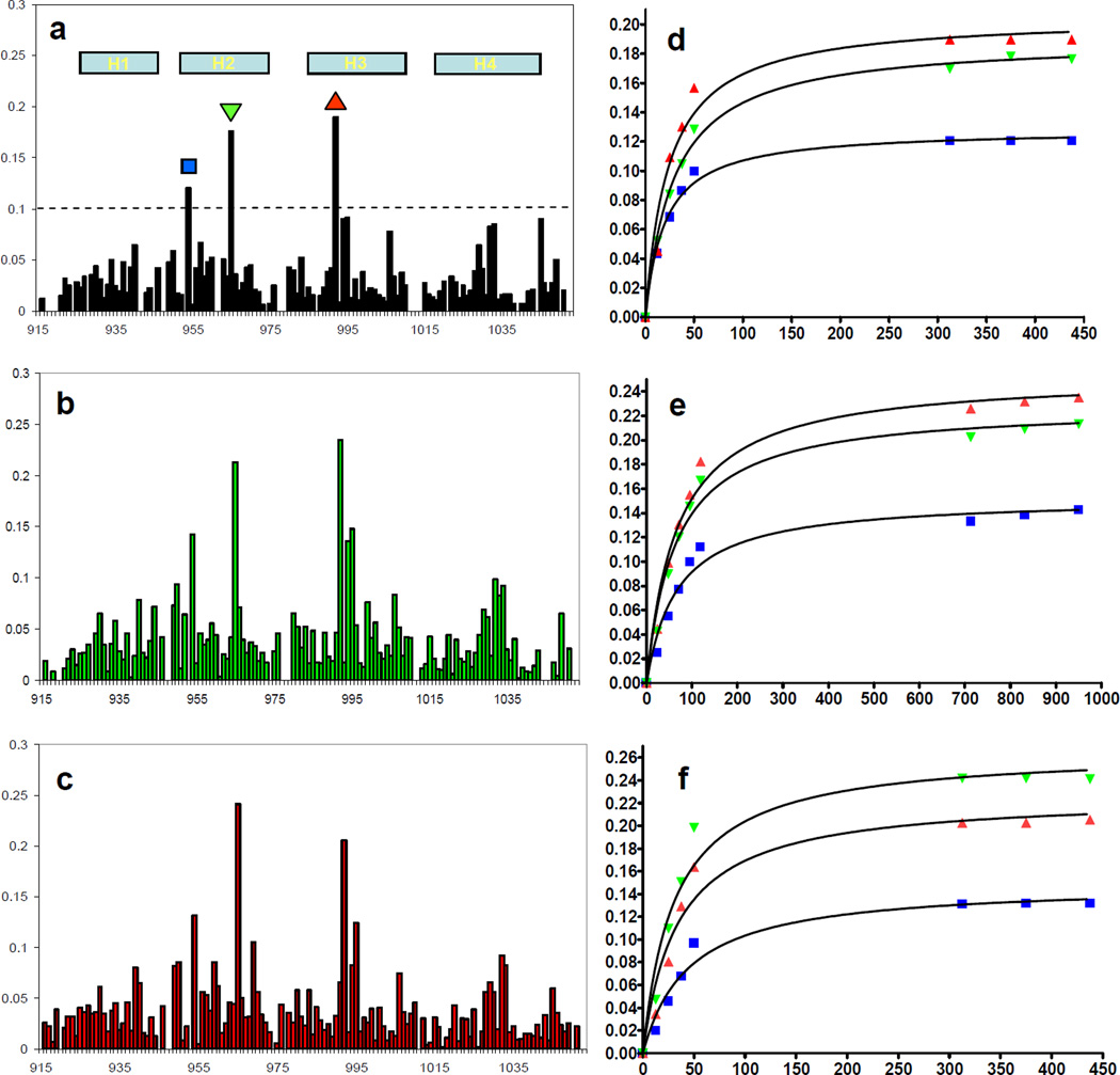
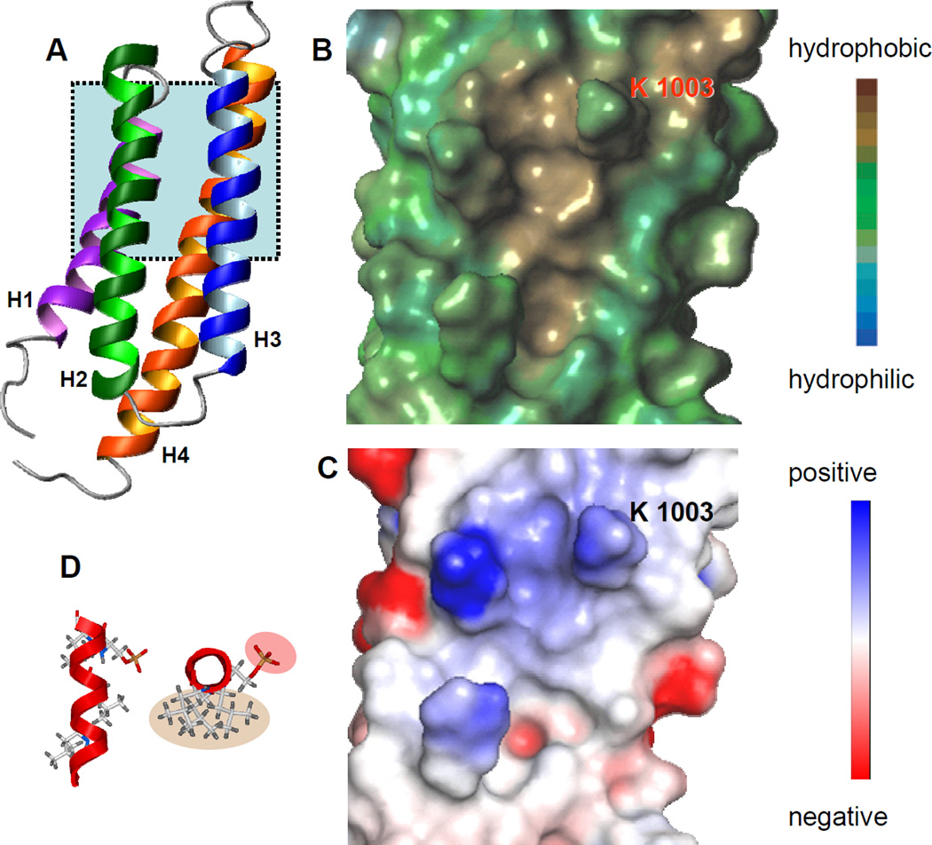
Cell migration is a dynamic process that requires the coordinated formation and disassembly of focal adhesions (FAs). Several proteins such as paxillin, focal adhesion kinase (FAK), and G protein-coupled receptor kinase-interacting protein 1 (GIT1) are known to play a regulatory role in FA disassembly and turnover. However, the mechanisms by which this occurs remain to be elucidated. Paxillin has been shown to bind the C-terminal domain of FAK in FAs, and an increasing number of studies have linked paxillin association with GIT1 during focal adhesion disassembly. It has been reported recently that phosphorylation of serine 273 in the LD4 motif of paxillin leads to an increased association with Git1 and focal adhesion turnover. In the present study, we examined the effects of phosphorylation of the LD4 peptide on its binding affinity to the C-terminal domain of FAK. We show that phosphorylation of LD4 results in a reduction of binding affinity to FAK. This reduction in binding affinity is not due to the introduction of electrostatic repulsion or steric effects but rather by a destabilization of the helical propensity of the LD4 motif. These results further our understanding of the focal adhesion turnover mechanism as well as identify a novel process by which phosphorylation can modulate intracellular signaling.
Figures
References
-
- Parsons JT. Focal adhesion kinase: the first ten years. J. Cell Sci. 2003;116:1409–1416. - PubMed
-
- Webb DJ, Parsons JT, Horwitz AF. Adhesion assembly, disassembly and turnover in migrating cells -- over and over and over again. Nat. Cell Biol. 2002;4:E97–E100. - PubMed
-
- Schlaepfer DD, Mitra SK, Ilic D. Control of motile and invasive cell phenotypes by focal adhesion kinase. Biochim. Biophys. Acta. 2004;1692:77–102. - PubMed
-
- McLean GW, Carragher NO, Avizienyte E, Evans J, Brunton VG, Frame MC. The role of focal-adhesion kinase in cancer - a new therapeutic opportunity. Nat. Rev. Cancer. 2005;5:505–515. - PubMed
Publication types
MeSH terms
Substances
Grants and funding
LinkOut - more resources
Full Text Sources
Molecular Biology Databases
Research Materials
Miscellaneous
