. 2007 Dec;4(12):e329.
doi: 10.1371/journal.pmed.0040329.

Detection of intracellular bacterial communities in human urinary tract infection

Affiliations

Detection of intracellular bacterial communities in human urinary tract infection

David A Rosen et al. PLoS Med. 2007 Dec.

Abstract

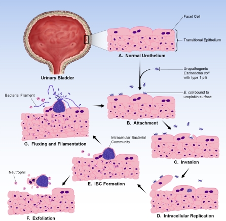
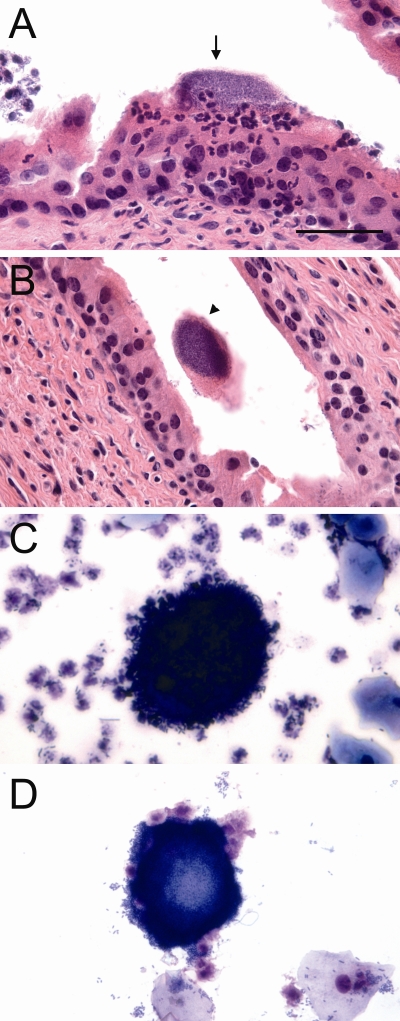
Background: Urinary tract infections (UTIs) are one of the most common bacterial infections and are predominantly caused by uropathogenic Escherichia coli (UPEC). While UTIs are typically considered extracellular infections, it has been recently demonstrated that UPEC bind to, invade, and replicate within the murine bladder urothelium to form intracellular bacterial communities (IBCs). These IBCs dissociate and bacteria flux out of bladder facet cells, some with filamentous morphology, and ultimately establish quiescent intracellular reservoirs that can seed recurrent infection. This IBC pathogenic cycle has not yet been investigated in humans. In this study we sought to determine whether evidence of an IBC pathway could be found in urine specimens from women with acute UTI.

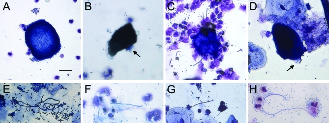
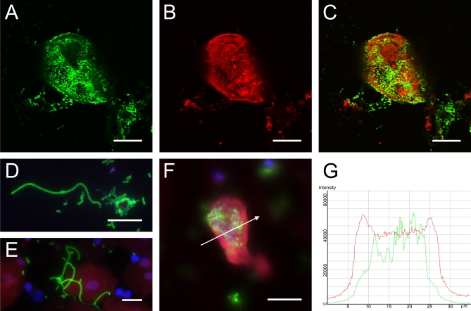
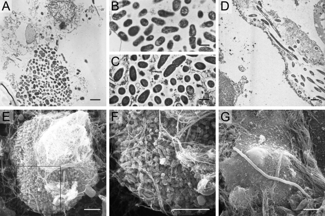
Methods and findings: We collected midstream, clean-catch urine specimens from 80 young healthy women with acute uncomplicated cystitis and 20 asymptomatic women with a history of UTI. Investigators were blinded to culture results and clinical history. Samples were analyzed by light microscopy, immunofluorescence, and electron microscopy for evidence of exfoliated IBCs and filamentous bacteria. Evidence of IBCs was found in 14 of 80 (18%) urines from women with UTI. Filamentous bacteria were found in 33 of 80 (41%) urines from women with UTI. None of the 20 urines from the asymptomatic comparative group showed evidence of IBCs or filaments. Filamentous bacteria were present in all 14 of the urines with IBCs compared to 19 (29%) of 66 samples with no evidence of IBCs (p < 0.001). Of 65 urines from patients with E. coli infections, 14 (22%) had evidence of IBCs and 29 (45%) had filamentous bacteria, while none of the gram-positive infections had IBCs or filamentous bacteria.

Conclusions: The presence of exfoliated IBCs and filamentous bacteria in the urines of women with acute cystitis suggests that the IBC pathogenic pathway characterized in the murine model may occur in humans. The findings support the occurrence of an intracellular bacterial niche in some women with cystitis that may have important implications for UTI recurrence and treatment.


Conflict of interest statement

Competing Interests: The authors have declared that no competing interests exist.

Figures

Figure 1
Figure 1. UPEC IBC Pathogenic Pathway Observed in the Murine Cystitis Model
The bladder urothelium (A) is a pseudostratified transitional epithelium lined by large facet cells. These cells have an apical asymmetric unit membrane containing uroplakins that help form the impermeable bladder barrier and also serve as receptors for UPEC. Bacteria introduced into the bladder adhere to the bladder surface via type 1 pili (B). Upon attachment, bacteria are able to invade (C) and replicate (D) within the facet cell cytoplasm. UPEC form large biofilm-like IBCs within these cells (E). Ultimately the bacteria flux out of their intracellular niche (G), some adopting a filamentous morphology; they then adhere to other host cells and re-enter the infectious cycle. During this process, infected urothelial cells are sloughed into the urine (F) and neutrophils are recruited to the site of infection.
Figure 2
Figure 2. Light Microscopy Findings of Potential IBCs and Filaments in Urines from Women with Cystitis
Light microscopy screening of urines from cystitis patients revealed biofilm-like collections of bacteria (A), potential intracellular bacterial communities (B–D) and filamentous bacteria (E–H). Many large biofilm-like collections of small, morphologically coccoid bacteria were found in cystitis urine samples. Dark-staining epithelial cells with potential IBCs were observed often with bacteria that appear to be protruding from within (arrows). Tangled collections and individual long filamentous bacteria were also found in several of the samples. Scale bar, 20 μm, applies to all photomicrographs.
Figure 3
Figure 3. Immunofluorescence Confirmation of IBCs and Filaments in Urines from Women with Cystitis
Urines from women with cystitis were stained with antibodies against E. coli (green) and uroplakin III (red). Confocal microscopy analysis revealed large collections of bacteria (A) and cells with partial membrane and cytoplasmic uroplakin staining (B). Merged images (C) show these bacteria to be intracellular. Filaments of the uropathogen over 20 μm in length were also observed in many of the urine samples (D and E). To quantify fluorescence, a slice was taken through the middle of an IBC (F) and fluorescent intensity was analyzed along a traversing line (arrow). A representative fluorescent intensity distribution profile (G) shows peaks of uroplakin (red) staining corresponding to the facet membrane and E. coli (green) staining localized intracellularly. Scale bars, 20 μm.
Figure 4
Figure 4. Electron Microscopy Findings in Urines from Women with Cystitis
TEM analysis of human cystitis urine specimens (A) revealed large collections of bacteria associated with nuclei and other cellular debris. These collections of bacteria from human urines (B) have similar morphology and organization as those recovered from intact murine intracellular bacterial communities (C). Bacteria and filaments were also observed intracellularly within exfoliated epithelial cells in a urine sample quickly fixed and analyzed from an E. coli cystitis patient (D). SEM analysis of cystitis urines deemed positive for IBCs and filaments captured large bacterial biofilm-like collections (E and F) composed of bacteria with a smaller, more coccoid morphology than typical E. coli. Long filaments were also captured by SEM (G). Scale bars, 2 μm (A and D), 1 μm (B and C), and 5 μm (E–G).
Figure 5
Figure 5. Mouse Trial and Comparison of Human and Mouse Urine
The E. coli isolate from a urine specimen positive for IBCs and filaments was inoculated into mice where it progressed through the IBC pathogenic cycle. Several IBCs were observed by H&E in the mouse bladder at 30 h postinfection (A, arrow). IBCs could also be seen exfoliated into the bladder lumen (B, arrowhead). Urine collected from mice at this time point were positive for IBCs (C). These IBCs were similar in morphology and size to those formed by the same E. coli isolate in the original human urine specimen (D). Scale bar, 50 μm, applies to all photomicrographs.

Comment in

References

    1. Griebling TL. Urinary Tract Infections in Women. In: Litwin MS, Saigal CS, editors. Urologic Diseases in America. US Department of Health and Human Services, Public Health Service, National Institutes of Health, National Institute of Diabetes and Digestive and Kidney Diseases; Washington (D. C.): US Government Printing Office; 2007. pp. 587–620. NIH Publication No. 07–5512, pp.
    1. Foxman B. Epidemiology of urinary tract infections: incidence, morbidity, and economic costs. Am J Med. 2002;113(Suppl 1A):5S–13S. - PubMed
    1. Hooton TM, Scholes D, Hughes JP, Winter C, Roberts PL, et al. A prospective study of risk factors for symptomatic urinary tract infection in young women. N Engl J Med. 1996;335:468–474. - PubMed
    1. Hooton TM, Stamm WE. Diagnosis and treatment of uncomplicated urinary tract infection. Infect Dis Clin North Am. 1997;11:551–581. - PubMed
    1. Ronald AR, Pattullo AL. The natural history of urinary infection in adults. Med Clin North Am. 1991;75:299–312. - PubMed

Publication types

MeSH terms