

Post-traumatic upper cervical subluxation visualized by MRI: a case report
- PMID: 18093309
- PMCID: PMC2253541
- DOI: 10.1186/1746-1340-15-20
Post-traumatic upper cervical subluxation visualized by MRI: a case report
Abstract
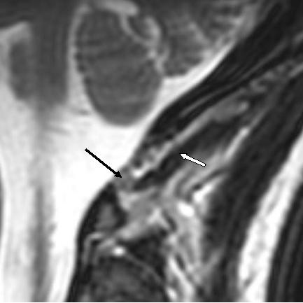
Background: This paper describes MRI findings of upper cervical subluxation due to alar ligament disruption following a vehicular collision. Incidental findings included the presence of a myodural bridge and a spinal cord syrinx. Chiropractic management of the patient is discussed.
Case presentation: A 21-year old female presented with complaints of acute, debilitating upper neck pain with unremitting sub-occipital headache and dizziness following a vehicular collision. Initial emergency department and neurologic investigations included x-ray and CT evaluation of the head and neck. Due to persistent pain, the patient sought chiropractic care. MRI of the upper cervical spine revealed previously unrecognized clinical entities.
Conclusion: This case highlights the identification of upper cervical ligamentous injury that produced vertebral subluxation following a traumatic incident. MRI evaluation provided visualization of previously undetected injury. The patient experienced improvement through chiropractic care.
Figures


References
-
- The Association of Chiropractic Colleges http://www.chirocolleges.org/missiont.html
-
- Benedetti PF, Fahr PM, Kuhns LR, Hayman LA. Imaging findings in spinal ligamentous injury. AJR. 2000;175:661–66. - PubMed
LinkOut - more resources
Full Text Sources
