. 2007 Dec 19:8:27.
doi: 10.1186/1471-2091-8-27.

Downregulation of COP9 signalosome subunits differentially affects the CSN complex and target protein stability

Affiliations

Downregulation of COP9 signalosome subunits differentially affects the CSN complex and target protein stability

Andreas Peth et al. BMC Biochem. .

Abstract

Background: The COP9 signalosome (CSN) is a conserved protein complex in eukaryotic cells consisting of eight subunits (CSN1 to CSN8). Recent data demonstrate that the CSN is a regulator of the ubiquitin (Ub) proteasome system (UPS). It controls substrate ubiquitination by cullin-RING Ub ligases (CRLs), a process that determines substrate specificity of the UPS. The intrinsic deneddylating activity localized to CSN5 as well as the associated kinases and deubiquitinating activity are involved in the regulatory function of CSN. The exact mechanisms are unclear. In this study we knocked down CSN1 (siCSN1), CSN3 (siCSN3) and CSN5 (siCSN5) by specific siRNA oligos permanently expressed in HeLa cells. The analysis and comparison of siRNA cells revealed differential impact of individual subunits on CSN structure and function.

Results: Permanent knockdowns of CSN1 and CSN3 led to a reduction of the subunits to approximately 40%, which is accompanied by a proportional decrease of the CSN holocomplex. In contrast, downregulation of CSN5 in HeLa cells reduced the CSN5 protein below 20% without significant effects on the remaining complex. The CRL component Rbx1 was characterized by accelerated proteolysis in siCSN1 and siCSN3 and also in siCSN5 cells, however, with lesser extent. Immunoprecipitated CSN complex from siCSN5 cells was less effective in phosphorylating c-Jun and p27. Accelerated degradation of c-Jun in siCSN5 cells was rescued by overexpression of CSN5 as well as of the deneddylation mutant CSN5D151N. Overexpression of CSN5 cannot rescue c-Jun destabilization in siCSN1.

Conclusion: There exists a coordinated downregulation of CSN subunits in the CSN1 and CSN3 knockdowns. The underlying regulatory mechanisms are obscure. CSN5 seems to possess a specific status in HeLa cells. Its reduction is not connected with coordinated downregulation of other subunits. CSN knockdowns confirm that the stabilization of the CRL component Rbx1 is a major CSN function. In addition, downregulation of CSN subunits influences the stability of important cellular regulators such as c-Jun and p27.


Figures
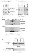
Figure 1
Figure 1
Comparison of HeLa cells permanently expressing siRNA oligos against CSN1, CSN3 or CSN5. (a) Western blots with lysates from HeLa cells permanently expressing siRNA oligos against GFP (siGFP cells = control cells), CSN1 (siCSN1 cells), CSN3 (siCSN3 cells) and CSN5 (siCSN5 cells) were performed using the anti-CSN5 and the anti-CSN8 antibody. RPN2 was analyzed as a loading control. The signals obtained with the anti-CSN5 and the anti-CSN8 antibodies were evaluated by densitometry. Values obtained in control cells were put to 100%. Similar blots with anti-CSN1 and anti-CSN3 antibodies were published recently [29]. 20S indicates the position of the 20S proteasome in the non-denaturing gels under our conditions. (b) Non-denaturing gel electrophoresis and blot with lysates from control cells (siGFP) and siCSN5 cells using the Phast-gel system. The same blot was probed with the anti-CSN5 antibody and the anti-CSN8 antibody. Whereas the anti-CSN5 antibody revealed a reduction to 23% as compared to the control, there was no decrease of the CSN complex observed with the anti-CSN8 antibody. (c) Glycerol gradient centrifugation of lysates from siGFP and siCSN5 cells. Aliquots of fractions 1 to 19 were probed by Western blotting using the anti-CSN5 and the anti-CSN8 antibody. Under these conditions the CSN complex sedimented into fractions 9 to 13. Free subunits were not detected. Under these conditions the 20S proteasome sedimented into fraction 5 to 9. (d) Control cells (siGFP) and siCSN5 cells were transfected with Flag-CSN5wt. The upper panels demonstrate Western blots with the anti-Cul1 antibody demonstrating two bands, deneddylated and mono-neddylated Cul1. There was significantly more mono-neddylated Cul1 in siCSN5 cells (vector) as compared to siGFP cells. Deneddylation was restored by overexpression of CSN5. The lower panel shows Western blots with the anti-CSN5 antibody visualizing the endogenous and the Flag-tagged exogenous CSN5 protein.
Figure 2
Figure 2
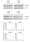
The CRL component Rbx1 is destabilized in CSN knockdown cells. (a) CHX chase experiments were performed with siGFP, siCSN1, siCSN3 and siCSN5 cells. After the indicated time cell lysates were analyzed by Western blotting using the anti-Rbx1 antibody. The α6 proteasomal subunit was probed as loading control. (b) Densitometric analysis of the Rbx1 signals from (a). The densitometric signal at 0 h was put to 100% and the percentage of Rbx1 signals was plotted against the indicated time.
Figure 3
Figure 3
The degradation of c-Jun is accelerated in CSN knockdown cells. (a) CHX chase experiments were performed with siGFP, siCSN1, siCSN3 and siCSN5 cells. After indicated time aliquots of cell lysates were analyzed by Western blotting using the anti-c-Jun antibody. The same samples were probed with the antibody against the 26S proteasome base subunit RPN2/S1 as a loading control. (b) CSN5 knockdown cells were transfected with CSN5wt as in Fig. 1d or with CSN5D151N and 24 h after transfection CHX chase experiments were carried out as in (a). The middle panel shows Western blots with the anti-Flag antibody visualizing the expressed CSN5wt or CSN5D151N proteins (c) CSN5wt was overexpressed in siCSN1 cells and after 24 h CHX experiments were performed as in (a). In all CHX experiments a band just above c-Jun appeared after 20 – 30 min, which cross-reacted with the anti-c-Jun antibody (in some blots it was cut off). This protein might be a modified c-Jun. The nature of this modification is currently unknown. Both the putative modified c-Jun as well as unmodified c-Jun disappeared during the experiment.
Figure 4
Figure 4
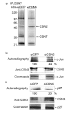
Phosphorylation of c-Jun and p27 by the CSN immunoprecipitated from siCSN5 cells. (a) Autophosphorylation of CSN subunits CSN2 and CSN7 with CSN immunoprecipitates from control cells (siGFP) and from siCSN5 cells. The CSN was precipitated with the anti-CSN7 antibody. The immunoprecipitate was incubated with 32P-γ-ATP, separated by SDS-PAGE and the dried gel was exposed to X-ray films. The autoradiography shows phosphorylation of CSN2 and CSN7 in the precipitate from siGFP as well as siCSN5 cells. Although same numbers of cells were used and the amount of the CSN complex is almost identical in control and siCSN cells, there was a slight increase of CSN2 and a decrease of CSN7 and overall phosphorylation with the CSN from siCSN5 cells. The exact reasons for these differences are not known at the moment. (b) Immunoprecipitates from siGFP or siCSN5 cells were used to phosphorylate c-Jun. The upper panel shows the phosphorylation of c-Jun by autoradiography demonstrating a decrease of c-Jun phosphorylation by more than 50% as estimated by densitometry. In the middle panel aliquots were analyzed by Western blotting using the anti-CSN3 antibody to control that equal amounts of the CSN were immunoprecipitated. The lower panel shows the Coomassie stain of c-Jun indicating that the same amounts of the protein were used for kinase assays. (c) The same experiments as performed with c-Jun (see b) were carried out with p27. As seen in the autoradiography the phosphorylation of p27 is significantly reduced with the CSN from siCSN5 cells as compared to the control.
Figure 5
Figure 5
Binding of p27 and of EB1 to the CSN determines the stability of the two cellular regulators. (a) Steady state levels of p27 were determined by Western blotting in siCSN1 and siGFP HeLa cells, which were synchronized as described before [14]. After reentry into G1 phase Western blots were performed with aliquots from the cytoplasm obtained after 0, 4 and 10 h. The cytoplasmic fraction was prepared as described using SOS protein as a marker for the cytoplasm [14]. As a loading control β-tubulin was analyzed. At the beginning of G1 phase the cell cycle inhibitor p27 was stabilized in the cytoplasm of CSN1 knockdown cells as compared to control cells (siGFP). (b) Purified CSN, recombinant GST-EB1 and increasing amounts of recombinant His-CSN5 were incubated for 30 min at 37°C. After incubation the mixture was directly analyzed by Western blotting (Input) or GST-pulldowns were performed and the precipitates were probed by anti-EB1 and anti-CSN3 antibodies. With increasing amounts of His-CSN5 less CSN complex was pulled down with GST-EB1 indicating that the binding between EB1 and the CSN was disturbed by CSN5. (c) The impact of the overexpression of CSN5wt or CSN5D151N mutant on EB1 steady state levels in HeLa cells. Transfection with the empty vector was used as control. Cells were lyzed 24 h after transfection and aliquots were analyzed by Western blotting using the anti-EB1 antibody (upper panel). The same lysates were tested with the anti-CSN5 antibody showing the endogenous and the Flag-tagged exogenous CSN5 protein (middle panel). The proteasome subunit RPN2/S1 was probed as a loading control (lower panel).

Similar articles

Cited by

References

    1. Bech-Otschir D, Kapelari B, Dubiel W. The COP9 Signalosome: Its Possible Role in the Ubiquitin System. In: Mayer R, Ciechanover A, Rechsteiner M, editor. Protein Degradation. 1: Ubiquitin and the Chemistry of Life. Weinheim , WILEY-VCH Verlag GmbH & Co. KGaA; 2005. pp. 348–369.
    1. Wei N, Deng XW. The COP9 signalosome. Annu Rev Cell Dev Biol. 2003;19:261–286. - PubMed
    1. Deng XW, Dubiel W, Wei N, Hofmann K, Mundt K, Colicelli J, Kato J, Naumann M, Segal D, Seeger M, Carr A, Glickman M, Chamovitz DA. Unified nomenclature for the COP9 signalosome and its subunits: an essential regulator of development. Trends Genet. 2000;16:202–203. - PubMed
    1. Maytal-Kivity V, Reis N, Hofmann K, Glickman MH. MPN+, a putative catalytic motif found in a subset of MPN domain proteins from eukaryotes and prokaryotes, is critical for Rpn11 function. BMC Biochem. 2002;3:28. - PMC - PubMed
    1. Verma R, Aravind L, Oania R, McDonald WH, Yates JR, 3rd, Koonin EV, Deshaies RJ. Role of Rpn11 metalloprotease in deubiquitination and degradation by the 26S proteasome. Science. 2002;298:611–615. - PubMed

Publication types

MeSH terms

Substances

LinkOut - more resources