Comment
. 2007 Dec 20;56(6):979-91.
doi: 10.1016/j.neuron.2007.11.013.

Structural analysis of the synaptic protein neuroligin and its beta-neurexin complex: determinants for folding and cell adhesion

Affiliations
Comment

Structural analysis of the synaptic protein neuroligin and its beta-neurexin complex: determinants for folding and cell adhesion

Igor P Fabrichny et al. Neuron. .

Abstract

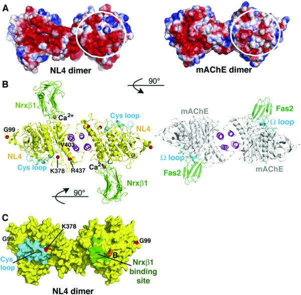
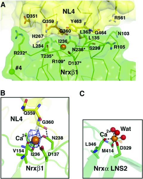
The neuroligins are postsynaptic cell adhesion proteins whose associations with presynaptic neurexins participate in synaptogenesis. Mutations in the neuroligin and neurexin genes appear to be associated with autism and mental retardation. The crystal structure of a neuroligin reveals features not found in its catalytically active relatives, such as the fully hydrophobic interface forming the functional neuroligin dimer; the conformations of surface loops surrounding the vestigial active center; the location of determinants that are critical for folding and processing; and the absence of a macromolecular dipole and presence of an electronegative, hydrophilic surface for neurexin binding. The structure of a beta-neurexin-neuroligin complex reveals the precise orientation of the bound neurexin and, despite a limited resolution, provides substantial information on the Ca2+-dependent interactions network involved in trans-synaptic neurexin-neuroligin association. These structures exemplify how an alpha/beta-hydrolase fold varies in surface topography to confer adhesion properties and provide templates for analyzing abnormal processing or recognition events associated with autism.


Figures

Figure 1
Figure 1. Sequence Conservation within the Neuroligins and Comparison with Human Liver Carboxylesterase and mAChE
The NL4 sequence displayed is that of the recombinant Gln44-Thr619 protein used for crystallization; the FLAG epitope is not shown. The positions for alternatively spliced inserts A and B are indicated by vertical arrowheads. The NL4 and mAChE numbering and their secondary structure elements are displayed above and below the alignment, respectively. The Cys-loop and loops L1 (α3 6,74 6,7, Thr332-Ile338 in NL4), L2 (α1 7,82 7,8, Asp381-Val390), L3 (β81 8,9, Gln477-Ser487) and L4 (α1 8,92 8,9, Gly503-Ser513) are indicated by gray bars under the alignment. The NL4 residues buried at the Nrxβ1 binding interface are shaded as gray boxes. The mAChE catalytic triad residues are indicated by asterisks; the two NL4 glycosylation sites by triangles; the residue pair involved in Ca2+ binding by filled circles; and the NL3/NL4 residues mutated in autism patients by filled squares. The LRE motif is indicated with a black bar above the sequences.
Figure 2
Figure 2. Overall View of the NL4 Structure
(A) (Left) The NL4 subunit is displayed with yellow β strands, orange α helices, green disulfide bridges, and an encircled vestigial catalytic triad. The GlcNAc moieties are visible; the N and C termini are labeled. (Right) The NL4 subunit, oriented as in (A), is colored according to temperature factor (B-factor values). The flexibility of the Cys-loop (orange) is evident. (B) Overall views of the NL4 dimer with the C termini pointing down (top) and with 90° rotation around the main axis (bottom). The four-helix bundle is displayed in magenta. The positions for splice sites A and B are indicated by green and red spheres, respectively. (C) Close-up view of the superimposed four-helix bundle regions in the NL4 (yellow, orange, and magenta) and mAChE (gray) dimers, showing the twist of the helical region at the subunit interface.
Figure 3
Figure 3. The NL4 Central Pocket Compared with the mAChE Active Center
(A) Close-up view of the tip of the Cys-loop (cyan) occluding access to the central pocket in NL4. The internal pocket, containing the phosphate, is displayed as a mesh surface. Residue Gly254 at the base of the nucleophilic elbow (β55,6 turn) is shown as a yellow sphere. (B) Close-up superimposition of the vestigial active center in NL4, rotated 90° from (A), and the active center in mAChE (rmsd, 0.3Å for 34 Cα atoms). The NL4 residues in the pocket are shown in yellow and those from the sealing Cys-loop in cyan. The mAChE residues in the pocket with Trp86 at the top are shown in white and those in the acyl pocket are shown in green. The NL4 Gly254 substitution to mAChE Ser203 is visible at the center. Bound phosphate (PO4) in NL4 mimics bound acetate (ACT) in mAChE. The phosphate oxygen atoms establish five hydrogen bonds with the main chain nitrogen atoms of Gly175, Ser176, and Ala255 (Gly121, Gly122, and Ala204 in mAChE) and the side chains of Ser176 and His489 (His447 in mAChE). The bond with Ala255 lies behind the figure plan and is not visible.
Figure 4
Figure 4. Comparison of the Surface Loops in NL4 and mAChE
(A) The Cys-loop (Cys110-Cys146; cf. Figure 1) in NL4 and (B) the U-loop (Cys69-Cys96) in mAChE are displayed in cyan, loops L1 (Thr332-Ile338 in NL4) in yellow, loops L2 (Asp381-Val390) in orange, loops L3 (Gln477-Ser487) in green, and loops L4 (Gly503-Ser513) in magenta. The Cys-loop conformation is stabilized by several polar and nonpolar interactions between Leu120, His121, Asp122, and Trp127 at the loop edge, and Ser176, Glu179, Lys378, Gly490, and Leu507, which surround the central pocket. Label AC on mAChE denotes the active center. Molecular surfaces of NL4 ([A], right) and mAChE ([B], right) show the occluded surface in NL4 and the accessible gorge entry, marked by the arrow, in mAChE. (C) Superimposition of the two Cys-loops (cyan and orange) and interacting loops L4 (magenta and violet) from the two NL4 subunits in the dimer. Residues that undergo large displacements are displayed. (D) Graph of differences in the loop sizes (in numbers of residues) of NL4, neurotactin (NRT), gliotactin (GLI), and glutactin (GLU) compared with mAChE (color codes as in [A]).
Figure 5
Figure 5. Electrostatic Potentials of the NL4 and mAChE Dimers and Comparison of the Dimeric Nrxβ1-NL4 and Fasciculin-mAChE Complexes
(A) The NL4 (left) and mAChE (right) dimers are oriented with their C termini pointing down (same as in Figure 2B, top). In each dimer, the two-fold symmetry axis places opposite faces of each subunit side by side. The encircled surface area is negatively charged in NL4, but not mAChE. This results in a continuum of electronegative surface potentials from one subunit face to the other face in the NL4 dimer, in contrast to the restricted localization of this potential to a single subunit face in the mAChE dimer. Electrostatic surface potentials are contoured at -3/+3 kT/e, where red describes a negative and blue a positive potential. (B) (Left) The Nrxβ1-NL4 complex with a yellow NL4, magenta four-helix bundle, cyan Cys-loops, and green Nrxβ1 molecules. The bound Nrxβ1 is oriented such that its curved β sheet is positioned in the prolongation of the curved long helix α4 7,8, which crosses the NL4 subunit. Red spheres denote the Gly99, Lys378, Val403, and Arg437 positions for mutations in NL3/NL4 of autism patients. (Right) The fasciculinmAChE complex with a gray mAChE, magenta bundle, cyan U-loops, and green fasciculins. Nrxβ1 (~25 kDa) and fasciculin (~7 kDa) bind on opposite faces of the NL4/mAChE dimer, through loops that connect b strands oriented perpendicular to the binding interfaces. The two complexes are oriented 90° compared with (A). (C) The NL4 dimer, oriented as in (A), is displayed with a yellow molecular surface, a cyan Cys-loop, and a green area buried by bound Nrxβ1. The position for splice site B in NL1 is displayed in orange, and the Gly99 and Lys378 positions for mutations in autism patients are displayed in red. The other mutations are not visible with this orientation.
Figure 6
Figure 6. Close-up View of the Ca2+-Mediated Nrxβ1-NL4 Complex Interface
(A) The Nrxβ1 molecular surface and key interacting side chains are displayed in green. The NL4 molecular surface is in yellow. The NL4 interacting side chains are in orange for the Ca2+-binding Gln359-Asn364 residues, and in yellow for the other residues. The Ca2+ ion is shown as an orange sphere central to the interface, and the position of splice site 4 in Nrxβ1 is shown as a green sphere. The asterisks denote Nrxβ1 residues whose mutation impairs binding to neuroligin (Graf et al., 2006). (B) Close-up view of the Ca2+ coordination at the complex interface (same orientation as in [A]). The omit difference electron density map (cyan) for Ca2+ is contoured at 3.5 s. Bonds with Nrxβ1 residues and likely bonds with NL4 residues are displayed as black and gray dotted lines, respectively. (C) Ca2+ coordination in the Nrxα LNS-2 domain structure (PDB accession code 2H0B; Sheckler et al., 2006) (same orientation as that of Nrxβ1 in [B]). The three coordinating water molecules are displayed as red spheres.

Comment in

Comment on

References

    1. Auld VJ, Fetter RD, Broadie K, Goodman CS. Gliotactin, a novel transmembrane protein on peripheral glia, is required to form the blood-nerve barrier in Drosophila. Cell. 1995;81:757–767. - PubMed
    1. Baker NA, Sept D, Joseph S, Holst MJ, McCammon JA. Electrostatics of nanosystems: application to microtubules and the ribosome. Proc. Natl. Acad. Sci. USA. 2001;98:10037–10041. - PMC - PubMed
    1. Blasi F, Bacchelli E, Pesaresi G, Carone S, Bailey AJ, Maestrini E. Absence of coding mutations in the X-linked genes neuroligin 3 and neuroligin 4 in individuals with autism from the IMGSAC collection. Am. J. Med. Genet. B. Neuropsychiatr. Genet. 2006;141:220–221. - PubMed
    1. Bolliger MF, Frei K, Winterhalter KH, Gloor SM. Identification of a novel neuroligin in humans which binds to PSD-95 and has a widespread expression. Biochem. J. 2001;356:581–588. - PMC - PubMed
    1. Botti SA, Felder CE, Sussman JL, Silman I. Electro-tactins: a class of adhesion proteins with conserved electrostatic and structural motifs. Protein Eng. 1998;11:415–420. - PubMed

Publication types