. 2007 Dec 20;56(6):1034-47.
doi: 10.1016/j.neuron.2007.11.014.

Potentiation of electrical and chemical synaptic transmission mediated by endocannabinoids

Affiliations

Potentiation of electrical and chemical synaptic transmission mediated by endocannabinoids

Roger Cachope et al. Neuron. .

Abstract

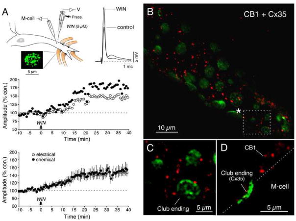
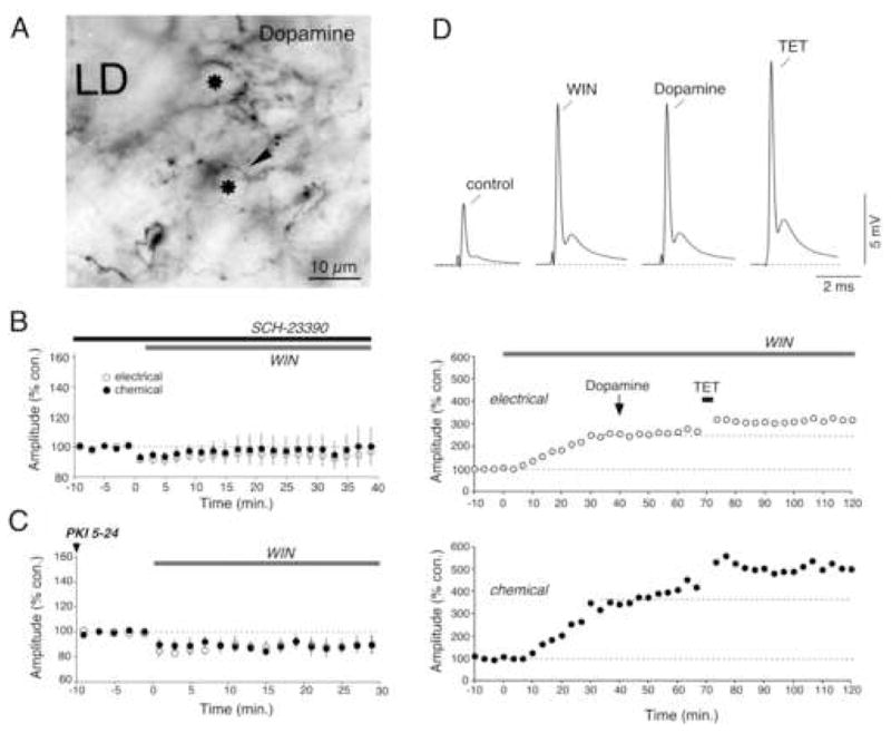
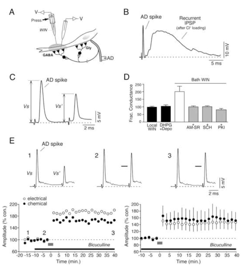
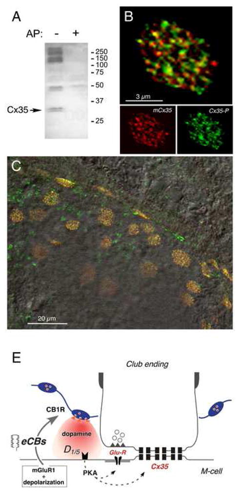
Endocannabinoids are well established as inhibitors of chemical synaptic transmission via presynaptic activation of the cannabinoid type 1 receptor (CB1R). Contrasting this notion, we show that dendritic release of endocannabinoids mediates potentiation of synaptic transmission at mixed (electrical and chemical) synaptic contacts on the goldfish Mauthner cell. Remarkably, the observed enhancement was not restricted to the glutamatergic component of the synaptic response but also included a parallel increase in electrical transmission. This effect involved the activation of CB1 receptors and was indirectly mediated via the release of dopamine from nearby varicosities, which in turn led to potentiation of the synaptic response via a cAMP-dependent protein kinase-mediated postsynaptic mechanism. Thus, endocannabinoid release can potentiate synaptic transmission, and its functional roles include the regulation of gap junction-mediated electrical synapses. Similar interactions between endocannabinoid and dopaminergic systems may be widespread and potentially relevant for the motor and rewarding effects of cannabis derivatives.


Figures

Figure 1
Figure 1. Cannabinoids evoke long-term potentiation of electrical and chemical transmission at Club endings
(A) Experimental arrangement. Electrical stimulation of the posterior branch of the VIIIth nerve (VIIIth nerve), where Club endings (Club endings, Mixed synapse) run, elicits a mixed excitatory postsynaptic potential (mixed EPSP) (B) Bath application of WIN 55,212-2 (WIN; 500 nM) results in a long-term enhancement of both components of the synaptic response. Top left, superimposed responses obtained 5 min before and 30 min after bath application of WIN (in all figures, traces represent the average of at least 10 responses). Top right, the antidromic spike (AD spike) height (a measure of M-cell input resistance) was slightly reduced. Bottom: time course of both components of the VIIIth nerve synaptic response (each point is the average of 10 single synaptic responses). (C) Averaged time course of the electrical and chemical components of the mixed EPSP after bath application of WIN (500 nM; n=8). (D) Pre-treatment with a combination of the CB1R antagonists AM251 and SR141716 (AM + SR; 4 μM ea) blocked WIN-evoked potentiation (n=8). (E) Application of AM and SR had no effect on the amplitude of the electrical or chemical components of the mixed EPSP (n=8).
Figure 2
Figure 2. Brief local activation of CB1R agonist is sufficient to trigger long-term potentiation of the mixed EPSP
(A) Experimental arrangement for local drug application in the vicinity of Club endings contacts on the distal portion of the lateral dendrite and labeling with anti-Cx35 antibodies. WIN (5 μM) was pressure ejected for 15 s. Traces represent the synaptic responses obtained 10 min before and 30 min after local application. Top: time course of both components of the mixed EPSP. Bottom: averaged time course of both components for 6 experiments with local WIN application. (B) Immunochemical analysis reveals the presence of CB1Rs in the vicinity of the M-cell lateral dendrite. Laser scanning confocal immunofluorescence in a double-immunolabeling experiment with anti-CB1 1-77 (red; Alexa fluor 594), and anti-Cx35/36 (green, Alexa fluor 488) antibodies. The image reconstructs a long stretch of the lateral dendrite (average of 3 confocal Z-sections, totaling 1.5 μm) showing abundant presence of CB1Rs (red) in the vicinity of the Club endings revealed by Cx35 labeling (green). (C) Higher magnification image of the area delimited by the dotted box in B, illustrating the close proximity of CB1Rs to Club endings. (D) Lateral view (average of 3 confocal Z-sections totaling 1.5 μm) shows labeling for CB1 in the immediate proximity of both the M-cell dendrite and a Club ending. The position of the M-cell membrane (dotted lines) was estimated from the DIC image of the section.
Figure 3
Figure 3. Dopamine mediates WIN-evoked potentiation of synaptic transmission at Club endings
(A) Dopaminergic innervation of the M-cell lateral dendrite (LD). Immunoreactive profiles obtained with an anti-dopamine antibody under Nomarski optics reveal the presence of numerous varicose fibers (arrow) lying between the Club endings, which appear in cross section (asterisk). (B) Averaged time course of both components of the mixed EPSP in 8 experiments in which WIN application followed pretreatment with the D1/5 antagonist SCH-23390 (50 μM). (C) Averaged time course for 5 experiments in which the PKA inhibitor PKI (500 μM) was intradendritically injected prior to bath application of WIN. (D) WIN-evoked potentiation occludes dopamine-evoked potentiation (10 mM). Top: Mixed EPSPs obtained at control (control) and after each experimental manipulation (WIN, Dopamine, TET) in one representative experiment. Below: time course of the amplitudes of the electrical and chemical components of the mixed EPSP. Bath application of WIN triggers a long-lasting potentiation of the mixed EPSP that occludes the effects of local dopamine application. In contrast, WIN-evoked potentiation did not occlude activity-dependent potentiation triggered by discontinuous high frequency stimulation of the VIIIth nerve (TET: burst of 6 pulses at 500 Hz, every 2 s over 4 minutes).
Figure 4
Figure 4. Endocannabinoids are released from the Mauthner cell lateral dendrite
(A) Immunochemical analysis reveals the presence of mGluR1 at Club endings. Laser scanning confocal immunofluorescence image obtained with polyclonal anti-mGlur1 (green, Alexa fluor 488); average of 11 confocal Z-sections totaling 5.5 μm. Club endings (arrowheads), unambiguously identified because of their larger size, exhibit punctate labeling for mGluR1. (B, C) Higher magnification images showing the distribution of mGluR1 at two Club endings. Note that the labeling is not restricted to the periphery of the contact, where chemical synapses are localized. (D–G) Both mGluR1 activation and dendritic depolarization are required for endocannabinoid release. (D) Time course of the electrical and chemical components of the mixed EPSP in experiments with local application of the mGluR I agonist DHPG (1 mM) in the vicinity of the M-cell dendrite (n=8). (E) Time course of experiments with dendritic depolarization of the M-cell (5 pulses, 5 s duration each, 90 nA of current, “depo”; n=6). (F) Combination of local application of DHPG and dendritic depolarization triggered long-lasting enhancement of the mixed EPSP (n=6). (G) CB1R antagonists AM and SR prevented the potentiation triggered by DHPG application and dendritic depolarization (n=6).
Figure 5
Figure 5. Endocannabinoid release is triggered by synaptic activity
(A) Repetitive stimulation (100 Hz) of the posterior VIIIth nerve provides both strong depolarization via gap junctions and glutamate release. (B) Repetitive stimulation of the posterior VIIIth nerve (100 Hz during 1 s × 5) evoked robust potentiation of both components of the mixed EPSP (n=5). (C) Pre-treatment with CB1R antagonists AM and SR (4 μM) blocked synaptic potentiation (n=5). (D) Intradendritic injections of THL (100 μM), a diacylglycerol lipase (DGL) inhibitor, prevented synaptic potentiation (n=6). (E) Dopamine D1/5R activation is required for synaptic potentiation triggered by repetitive stimulation. Pretreatment with the D1/5 antagonist SCH-23390 (50 μM) blocked synaptic potentiation (n=5).
Figure 6
Figure 6. Inhibitory synaptic transmission to the M-cell in the vicinity of Club endings is not modified by CB1R activation
(A) Experimental arrangement. The cartoon illustrates the relative distribution of inhibitory interneurons on the M-cell soma (mostly glycinergic) and lateral dendrite (mostly GABAergic), which are recurrently activated by antidromic stimulation (AD). (B) Cl loading of the M-cell reveals the presence of a powerful recurrent inhibitory synaptic potential in response to antidromic stimulation (AD spike). (C) The strength of the feed-back inhibition was quantified by determining the fractional conductance (expressed as % control). (D) Neither local application of WIN (Local WIN) nor local release of endocannabinoids triggered by DHPG and dendritic depolarization (DHPG+Depo) affected the strength of recurrent inhibition. In contrast, bath application of WIN (Bath WIN) led to a marked increase in recurrent inhibition suggesting that inhibitory synapses in the soma are sensitive to endocannabinoid modulation. This enhancement was prevented by application of CB1R antagonists AM and SR, the dopamine D1/5R antagonist SCH-23390 and the PKA blocker PKI. (E) Bath application of the GABAA receptor antagonist bicuculline (75 μM) prior to repetitive stimulation (100 Hz) of the posterior VIIIth nerve does not prevent the induction of activity-dependent synaptic potentiation. Bottom left: time course of the mixed EPSP from a single experiment during baseline recordings following repetitive stimulation of the posterior VIIIth nerve (gray bar). Note the lack of modification in baseline following the application of bicuculline and prior to repetitive stimulation. Top: responses to antidromic stimulation of the M-cell axon obtained at the time points 1 (−13 min), 2 (−4 min.) and 3 (40 min.) indicated in the graph. Bicuculline led to a suppression of recurrent inhibition at time points 2 and 3, indicated by the increase in the amplitude of the second antidromic action potential (bars at the second antidromic spikes in 2 and 3 indicate the amplitude prior to drug application). Bottom right: averaged time course of 5 experiments in which pre-treatment with bicuculline did not prevent the potentiation triggered by repetitive stimulation at 100Hz.
Figure 7
Figure 7. CB1Rs are closely associated to dopaminergic fibers
(A) Laser scanning confocal immunofluorescence with double-immunolabeling by polyclonal anti-CB1-CT (red, Alexa fluor 594) and monoclonal anti-TH (green; Alexa fluor 488) antibodies. Image is the average of 31 confocal Z-sections (totaling 6.2 μm). (B) Magnification of the boxed area from A illustrates close association between CB1Rs and dopaminergic fibers, compatible with presynaptic localization of CB1Rs on the membrane of the varicosities (image is the average of 9 Z-sections, totaling 1.8 μm).
Figure 8
Figure 8. PKA phosphorylates connexin 35 in Club endings
(A) Polyclonal anti-Cx35/P110 antibody recognizes Cx35 as a pair of bands at 32–33 kDa (arrow, left lane, AP-) in membranes from a small area of goldfish hindbrain where M-cells are located. Labeling is lost in membranes digested with alkaline phosphatase (right lane, AP+). The high molecular weight phospho-proteins labeled non-specifically by the antibody are not associated with Cx35 (Kothmann et al., 2007). 50 μg/lane crude membrane protein. (B) Laser scanning confocal immunofluorescence of a single Club ending with double-labeling by polyclonal anti-Cx35/P110 (green, Alexa fluor 488), and monoclonal anti-Cx35/36 (mCx35; red, Alexa fluor 594) antibodies. Top: superimposition of individual mCx35 and Cx35-P images (bottom), average of 3 confocal Z-sections (totaling 1.5 μm). (C) Lower magnification image showing extensive co-localization at individual Club endings in a section of the M-cell lateral dendrite. A DIC image of this region is superimposed. (D) Model for endocannabinoid-mediated potentiation of electrical and chemical synaptic transmission at Club endings. Synaptic activity leads to mGluR activation paired with postsynaptic membrane depolarization, triggering endocannabinoid (eCB) release from the postsynaptic M-cell dendrite, which activates CB1Rs on dopaminergic fibers. CB1R activation leads to dopamine release which, by activating postsynaptic D1/5 receptors, increases PKA activity responsible for simultaneous potentiation of electrical (Cx35) and glutamatergic (GluR) synaptic transmission.

Comment in

References

    1. Alger BE. Retrograde signaling in the regulation of synaptic transmission: focus on endocannabinoids. Prog Neurobiol. 2002;68:247–286. - PubMed
    1. Bash R, Rubovitch V, Gafni M, Sarne Y. The stimulatory effect of cannabinoids on calcium uptake is mediated by Gs GTP-binding proteins and cAMP formation. Neurosignals. 2003;12:39–44. - PubMed
    1. Bonhaus DW, Chang LK, Kwan J, Martin GR. Dual activation and inhibition of adenylyl cyclase by cannabinoid receptor agonists: evidence for agonist-specific trafficking of intracellular responses. J Pharmacol Exp Ther. 1998;287:884–888. - PubMed
    1. Brandes RP, Popp R, Ott G, Bredenkotter D, Wallner C, Busse R, Fleming I. The extracellular regulated kinases (ERK) 1/2 mediate cannabinoid-induced inhibition of gap junctional communication in endothelial cells. Br J Pharmacol. 2002;136:709–716. - PMC - PubMed
    1. Brenowitz SD, Regehr WG. Associative short-term synaptic plasticity mediated by endocannabinoids. Neuron. 2005;45:419–431. - PubMed

Publication types

MeSH terms

Substances

LinkOut - more resources