

Relaxation effects in the quantification of fat using gradient echo imaging
- PMID: 18093781
- PMCID: PMC2386876
- DOI: 10.1016/j.mri.2007.08.012
Relaxation effects in the quantification of fat using gradient echo imaging
Abstract
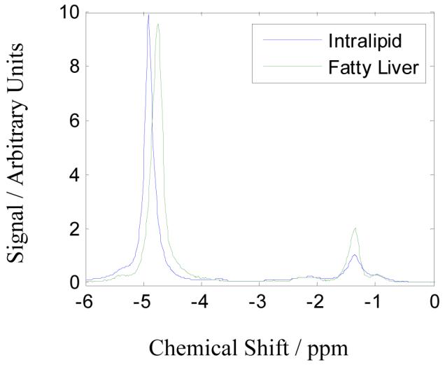
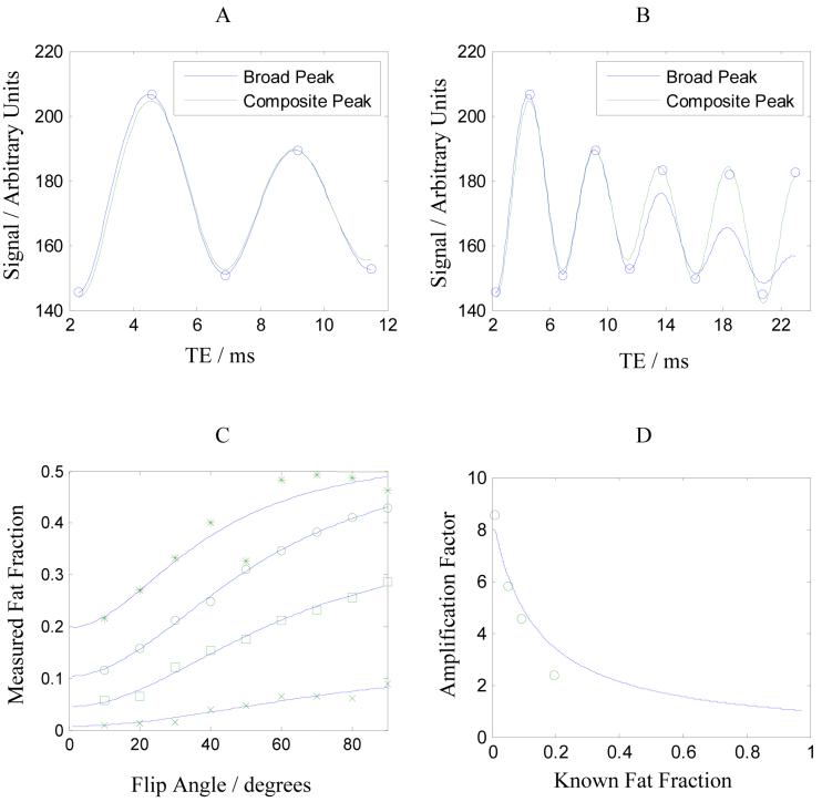
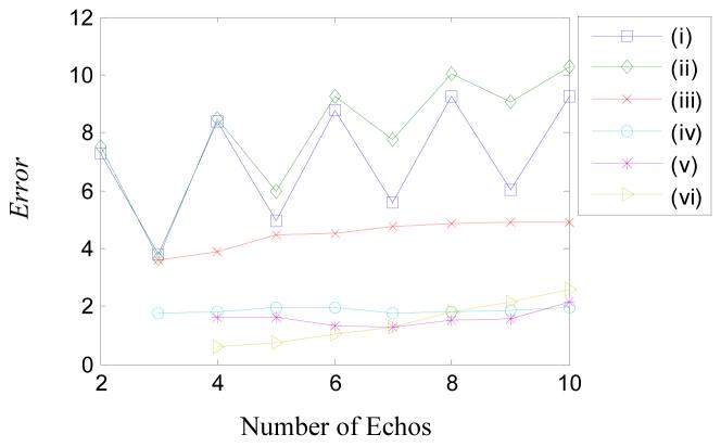
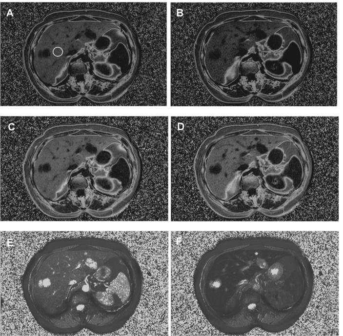
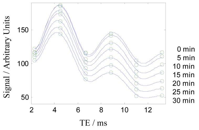
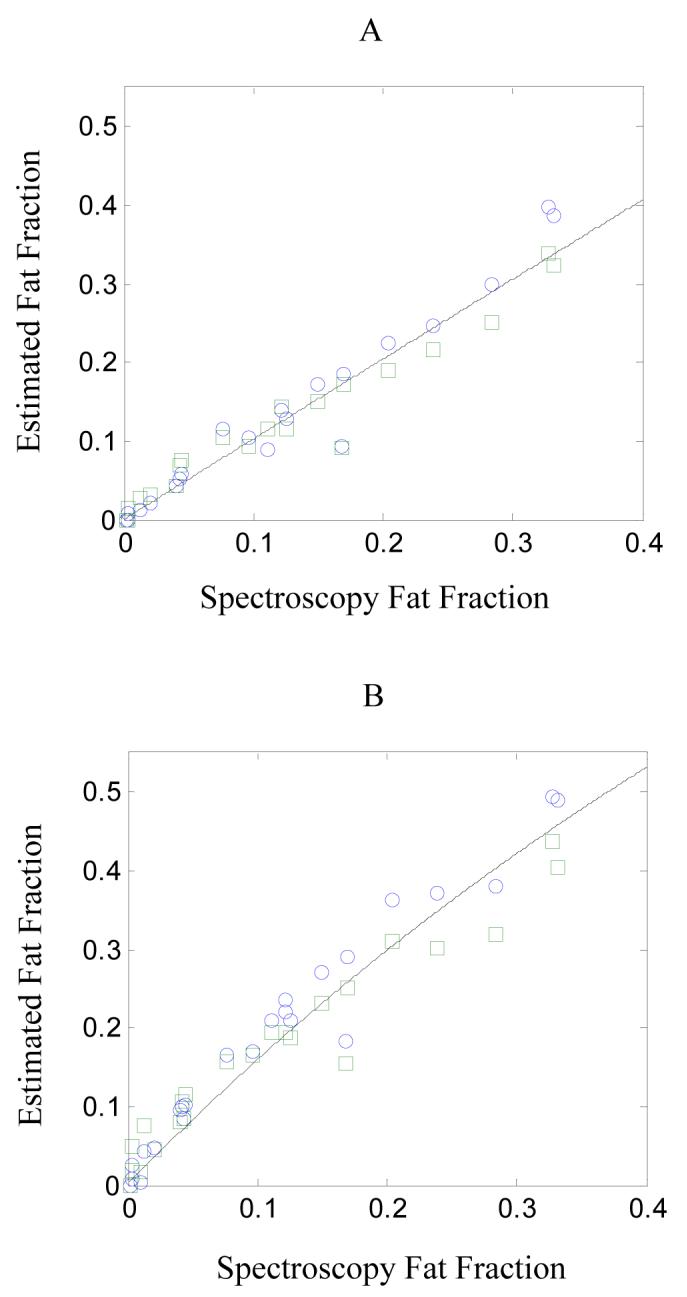
Quantification of fat has been investigated using images acquired from multiple gradient echoes. The evolution of the signal with echo time and flip angle was measured in phantoms of known fat and water composition and in 21 research subjects with fatty liver. Data were compared to different models of the signal equation, in which each model makes different assumptions about the T1 and/or T2* relaxation effects. A range of T1, T2*, fat fraction and number of echoes was investigated to cover situations of relevance to clinical imaging. Results indicate that quantification is most accurate at low flip angles (to minimize T1 effects) with a small number of echoes (to minimize spectral broadening effects). At short echo times, the spectral broadening effects manifest as a short apparent T2 for the fat component.
Figures

References
-
- Dixon WT. Simple Proton Spectroscopic Imaging. Radiology. 1984;153:189. - PubMed
-
- Glover GH. Multipoint Dixon Technique for Water and Fat Proton and Susceptibility Imaging. J Magn Reson Imag. 1991;1:521. - PubMed
-
- Yeung HN, Kormos DW. Separation of true fat and water images by correcting magnetic field inhomogeneity in situ. Radiology. 1986;159:783. - PubMed
-
- Xiang QS. Two-point water-fat imaging with partially-opposed-phase (POP) acquisition: an asymmetric Dixon method. Magn Reson Med. 2006;56:572. - PubMed
-
- Pauly J. Course notes on ‘Dixon Reconstruction’. http://www.stanford.edu/class/ee369c/notes/dixon.pdf.
Publication types
MeSH terms
Substances
Grants and funding
LinkOut - more resources
Full Text Sources
Other Literature Sources
Medical
