. 2008 Feb;20(2):443-52.
doi: 10.1016/j.cellsig.2007.11.005. Epub 2007 Nov 17.

Differential mediation of the Wnt canonical pathway by mammalian Dishevelleds-1, -2, and -3

Affiliations

Differential mediation of the Wnt canonical pathway by mammalian Dishevelleds-1, -2, and -3

Yi-Nan Lee et al. Cell Signal. 2008 Feb.

Abstract

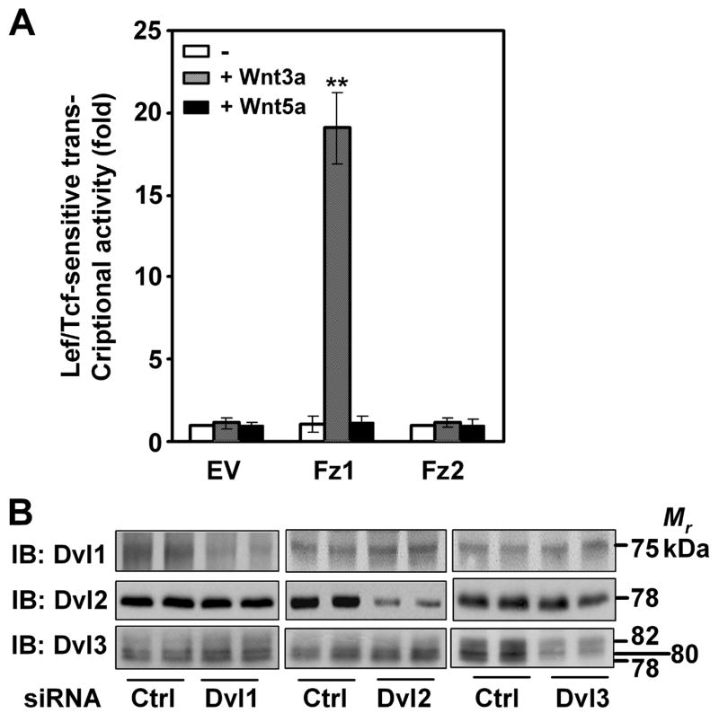
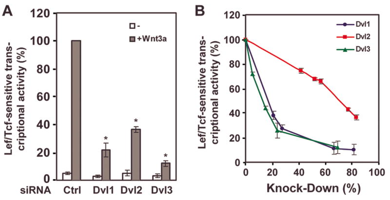
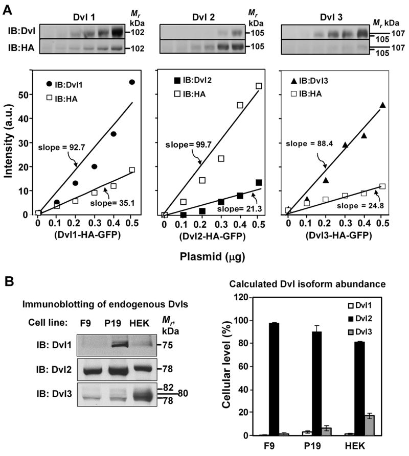
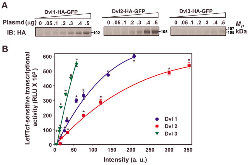
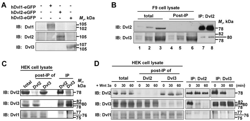
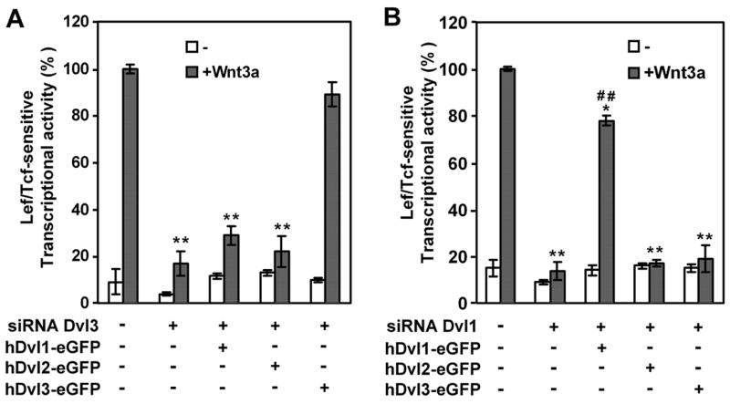
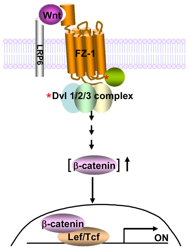
In the Drosophila, a single copy of the phosphoprotein Dishevelled (Dsh) is found. In the genomes of higher organism (including mammals), three genes encoding isoforms of Dishevelled (Dvl1, Dvl2, and Dvl3) are present. In the fly, Dsh functions in the Wnt-sensitive stabilization of intracellular beta-catenin and activation of the Lef/Tcf-sensitive transcriptional response known as the Wnt "canonical" pathway. In the current work we explore the expression of Dishevelleds in mammalian cells and provide an estimate of the relative cellular abundance of each Dvl. In mouse F9 cells, all three Dvls are expressed. Dvl2 constitutes more than 95% of the total pool, the sum of Dvl1 and Dvl3 constituting the remainder. Similarly, Dvl2 constitutes more than 80% of the Dvl1-3 pool in mouse P19 and human HEK 293 cells. siRNA-induced knock-down of individual Dvls was performed using Wnt3a-sensitive canonical pathway in F9 cells as the read-out. Activation of the canonical signaling pathway by Wnt3a was dependent upon the presence of Dvl1, Dvl2, and Dvl3, but to a variable extent. Wnt3a-sensitive canonical transcription was suppressible, by knock-down of Dvl1, Dvl2, or Dvl3. Conversely, the overexpression of any one of the three Dvls individually was found to be capable of promoting Lef/Tcf-sensitive transcriptional activation, in the absence of Wnt3a, i.e., overexpression of Dvl1, Dvl2, or Dvl3 is Wnt3a-mimetic. Graded suppression of individual Dvl isoforms by siRNA was employed to test if the three Dvls could be distinguished from one another with regard to mediation of the canonical pathway. Canonical signaling was most sensitive to changes in the abundance of either Dvl3 or Dvl1. Changes in expression of Dvl2, the most abundant of the three isoforms, resulted in the least effect on canonical signaling. Dvl-based complexes were isolated by pull-downs from whole-cell extracts with isoform-specific antibodies and found to include all three Dvl isoforms. Rescue experiments were conducted in which depletion of either Dvl3 or Dvl1 suppresses Wnt3a activation of the canonical pathway and the ability of a Dvl isoform to rescue the response evaluated. Rescue of Wnt3a-stimulated transcriptional activation in these siRNA-treated cells occurred only by the expression of the very same Dvl isoform depleted by the siRNA. Thus, Dvls appear to function cooperatively as well as uniquely with respect to mediation of Wnt3a-stimulated canonical signaling. The least abundant (Dvl1, 3) plays the most obvious role, whereas the most abundant (Dvl2) plays the least obvious role, suggesting that individual Dvl isoforms in mammals may operate as a network with some features in common and others rather unique.


Figures

Figure 1
Figure 1. Fz1-expressing mouse totipotent F9 teratocarcinoma cells display canonical activation by Wnt3a and express Dv1, Dvl2, and Dvl3
A, mouse F9 cells transfected to express Fz1 or Fz2 or empty vector (EV) were treated for 6 hr without or with purified Wnt3a (10 ng/ml) or Wnt5a (50 ng/ml) and the activation of the Wnt-sensitive canonical pathway monitored by the use of the Lef/Tcf-sensitive luciferase gene reporter. Data presented are mean values ± S.E.M. from three or more independent experiments. **, p ≤ 0.001 for the difference from control. B, Whole cell lysates from prepared F9 cells either untreated (Control) or treated with siRNA targeting either mDvl1, or mDvl2, or mDvl3. Samples of the lysates were subjected to SDS-PAGE and transfer of the resolved proteins to nitrocellulose blots. The blots were stained with primary antibodies against either Dvl1, or Dvl2, or Dvl3. A blot, representative of three or more individual, separate experiments is displayed. The tabulated data from these multiple experiments are presented as mean values ± S.E.M. from three or more independent experiments. **, p ≤ 0.01 for the difference from control can be found in Table 1.
Figure 2
Figure 2. siRNA-induced knockdown of Dvl1, Dvl2, or Dvl3 attenuated Wnt3a-stimulated canonical signaling
Mouse F9 cells expressing Fz1were treated with maximal (panel A) or variable (panel B) amounts of siRNAs targeting one of the three Dvls (Dvl1, Dvl2, or Dvl3) and the activation of the Wnt-sensitive canonical pathway in response to Wnt3a (6 hr) and monitored by the use of the Lef/Tcf-sensitive luciferase gene reporter. The assay of the canonical pathway was performed at 6 hr following stimulation without or with purified Wnt3a. Data presented are mean values ± S.E.M. from three or more independent experiments. *, p ≤ 0.01 for the difference from control. In panel A, the data are plotted as % of maximal Wnt3a-stimulated canonical response versus treatment with a maximal amount of siRNA reagent. In panel B, the data are plotted as “% of maximal” of the Wnt3a-sensitive canonical response versus the extent of knockdown in Dvl isoform protein produced by treatment increasing amounts of siRNA targeting a given Dvl isoform.
Figure 3
Figure 3. Determination of the relative abundance of Dvl1, Dvl2, and Dvl3 in mouse F9, mouse P19, and human HEK 293 cells
A, mouse F9 cells expressing Fz1 were co-transfected without or with increasing amounts of plasmid DNA (expression vector) harboring a single Dvl isoform tagged with both HA and GFP at the C-terminus of the molecule, as described. After 48 hr, the F9 cells were disrupted and sample of whole-cell lysates was subjected to SDS-PAGE and transfer of the resolved proteins to nitrocellulose blots that were then stained with primary antibodies against either Dvl1, or Dvl2, Dvl3, or the HA-antigen tag. A set of representative blots obtained from exogenously expressed Dvl isoforms (upper panels) and plots of expression of these isoforms (lower panels). B, analyses of the relative abundance of Dvl1, Dvl2, and Dvl3 in F9, P19, and HEK 293 cells. Samples (0.1 mg protein/SDS-PAGE lane) of whole-cell lysates were subjected to SDS-PAGE and transfer of the resolved proteins to nitrocellulose blots. The blots were stained with primary antibodies against Dvl1, or Dvl2, or Dvl3. A set of representative blots of endogenously expressed Dvl isoforms is displayed shown (left panels). These data were analyzed and the relative amount of Dvl isoform expressed in each cell calculated (right panel). *, p ≤ 0.01; **, p ≤ 0.001 for the difference from Dvl1 expression.
Figure 4
Figure 4. Effects of “over”expression of Dvl1, Dvl2, or Dvl3 on the activation of Lef/Tcf-sensitive transcriptional activation, in the absence of Wnt3a stimulation
Fz1-expressing F9 cells were transiently co-transfected with SuperX8TOPFLASH reporter gene in the absence or presence of increasing amounts of input plasmid DNA (expression vector) harboring either Dvl1, or Dvl2, or Dvl3 which had been tagged on the C-terminus with HA. After 48 hr, the cells were disrupted and whole-cell lysates prepared. A, sample of whole-cell lysates was subjected to SDS-PAGE and transfer of the resolved proteins to nitrocellulose blots. The blots were stained with primary anti-HA antibody and their relative levels of expression compared. B, the activation of the Lef/Tcf-sensitive transcription was assayed by using SuperX8TOPFLASH luciferase gene reporter. The luciferase reporter activities were plotted versus the amount of immunoreactive staining for the HA-tag (in arbitrary unit; a.u.) quantified in the blots obtained from the same cell lysates. Data presented are mean values ± S.E.M. from three or more independent experiments. *, p ≤ 0.01 for the difference from the “Control” cells that were treated with empty vector that lacks a Dvl isoform cDNA.
Figure 5
Figure 5. Dvl-based complexes are heterogeneous with respect to Dvl isoforms
A, Dvl isoform-specific antibodies were tested for cross reactivity by immunoblotting of whole-cell lysates obtained from cell “over”expressing a single Dvl isoform. Immunoblots of cell lines that were “over”expressing a single Dvl isoform then were stained with individual antibodies against each of the Dvl isoforms. B, immune precipitation-based “pull-downs” from whole-cell lysates of F9 cells were performed using antibodies that recognize only Dvl2. The Dvl-based complexes isolated in the pull-downs were subjected to SDS-PAGE and the resolved proteins transferred to nitrocellulose blots. The blots were stained with antibodies specific for each Dvl isoform, seeking to detect Dvl1, DVl2, and Dvl3 that may be present in the complex. C, immune precipitation-based “pull-downs” from whole-cell lysates of HEK293 cells were performed using antibodies that recognize only either Dvl2 or Dvl3. Whole-cell lysates (“total”), pull-down complexes (“IP”) as well as supernatant fraction following the immune precipitation reaction (“post-IP”) were subjected to SDS-PAGE and the resolved proteins transferred to nitrocellulose blots. The blots were stained for each Dvl isoform. D, HEK293 cells expressing Fz1 were treated without and with purified Wnt3a for up to 60 min and the composition of Dvl2-based as well as Dvl3-based complexes analyzed for the presence of all three isoforms in the complex performed by immunoblotting. Immune precipitation-based “pull-downs” were performed using antibodies that recognize either Dvl2 or Dvl3. Whole-cell lysates (total), pull-down complexes (IP) as well as supernatant from post-immunoprecipitated fraction (post-IP) were subjected to SDS-PAGE and the resolved proteins transferred to nitrocellulose blots and stained for each Dvl isoform. The data presented in these panels are blots representative of three or more independent analyses for each protocol. The data from each of the experiments are in agreement.
Figure 6
Figure 6. Rescue of Wnt3a-stimulated Lef/Tcf-sensitive transcriptional activation of Dvl3-or Dvl1-depleted mouse F9 cells using exogenous expression of hDvl1, hDvl2, or hDvl3
Fz1-expressing mouse F9 cells were treated with siRNA targeting either mouse Dvl1 (A) or Dvl3 (B). At 24 hr post addition of the siRNA, the cells were co-transfected with an expression vector harboring hDvl1, hDvl2, or Dvl3 which were tagged with eGFP at its C-terminus as well as with the SuperX8TOPFLASH luciferase reporter gene construct and the culture extended for another 24 hr. The assay of the Wnt3a-sensitive canonical pathway was performed by the assay of the SuperX8TOPFLASH luciferase reporter at 6 hr following stimulation without or with purified Wnt3a. A, rescue of Wnt3a-stimulated activation of the canonical pathway in Dvl3-depleted F9 cells by exogenous expression of human Dvl1, Dvl2, or Dvl3. B, rescue of Wnt3a-stimulated activation of the canonical pathway in Dvl1-depleted F9 cells by exogenous expression of human Dvl1, Dvl2, or Dvl3. The human Dvl isoforms are resistant to suppression by siRNAs targeting the mouse homologues. Data presented are mean values ± S.E.M. from three or more independent experiments. *, p ≤ 0.01; **, p ≤ 0.001 for the difference from control, +Wnt3a group. ##, ≤ 0.001 for the difference from group treated with siRNA alone.
Figure 7
Figure 7. A schematic presentation of Dvl complex containing Dvl1, Dvl2 and Dvl3 in Frizzled-1/β-catenin signaling pathway
*, denotes “activation” in response to Wnt3a.

Similar articles

Cited by

References

    1. Mikels AJ, Nusse R. Wnts as ligands: processing, secretion and reception. Oncogene. 2006;25:7461–7468. - PubMed
    1. Wang HY, Liu T, Malbon CC. Structure-function analysis of Frizzleds. Cell Signal. 2006;18:934–941. - PubMed
    1. Kikuchi A, Yamamoto H, Kishida S. Multiplicity of the interactions of Wnt proteins and their receptors. Cell Signal. 2007;19:659–671. - PubMed
    1. Malbon CC. G proteins in development. Nat Rev Mol Cell Biol. 2005;6:689–701. - PubMed
    1. Cadigan KM, Nusse R. Wnt signaling: a common theme in animal development. Genes Dev. 1997;11:3286–3305. - PubMed

Publication types

MeSH terms