. 2008 Mar;191(1):24-30.
doi: 10.1016/j.jmr.2007.11.018. Epub 2007 Dec 3.

Continuity conditions and torsion angles from ssNMR orientational restraints

Affiliations

Continuity conditions and torsion angles from ssNMR orientational restraints

S Achuthan et al. J Magn Reson. 2008 Mar.

Abstract

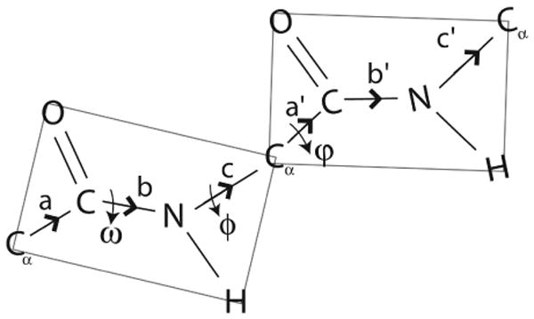
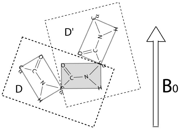
The backbone torsion angle pair (varphi,psi) at each amino acid of a polypeptide is a descriptor of its conformation. One can use chemical shift and dipolar coupling data from solid-state NMR PISEMA experiments to directly calculate the torsion angles for the membrane-spanning portion of a protein. However, degeneracies inherent in the data give rise to multiple potential torsion angles between two adjacent peptide planes (a diplane). The molecular backbone structure can be determined by gluing together the consecutive diplanes, as in the PIPATH algorithm [T. Asbury, J.R. Quine, S. Achuthan, J. Hu, M.S. Chapman, T.A. Cross, R. Bertram, PIPATH: an optimized alogrithm for generating alpha-helical structures from PISEMA data, J. Magn. Reson. 183 (2006) 87-95.]. The multiplicities in torsion angles translate to multiplicities in diplane orientations. In this paper, we show that adjacent diplanes can be glued together to form a permissible structure only if they satisfy continuity conditions, described quantitatively here. These restrict the number of potential torsion angle pairs. We rewrite the torsion angle formulas from [J.R. Quine, M.T. Brenneman, T.A. Cross, Protein structural analysis from solid-state NMR-drived orientational constraints, Biophys. J. 72 (1997) 2342-2348.] so that they automatically satisfy the continuity conditions. The reformulated torsion angle formulas have been applied recently in the PIPATH algorithm [T. Asbury, J.R. Quine, S. Achuthan, J. Hu, M.S. Chapman, T.A. Cross, R. Bertram, PIPATH: an optimized alogrithm for generating alpha-helical structures from PISEMA data, J. Magn. Reson. 183 (2006) 87-95.] and will be helpful in other applications in which diplane gluing is used to construct a protein backbone model.


Figures

Figure 1
Figure 1
A typical PISEMA powder pattern bounded by primary and reflected ellipses (PISEMA ellipses) and a small extra-elliptical triangle (PISEMA triangle) near Q. The experimental data (σ,|ν|2) fall within the shaded regions A-E. The number of degeneracies associated with each region of the powder pattern is as follows: regions A and B (4-fold degeneracy), regions C and D (8-fold degeneracy) and region E (12-fold degeneracy). We assume that the angle between the N-H bond vector and σ33 is α = 0° and β = 17° [17].
Figure 2
Figure 2
Torsion angles and bond vectors of a diplane. The bond vectors of the second peptide plane have primed superscripts.
Figure 3
Figure 3
An oriented structure consisting of three peptide planes obtained by gluing two oriented diplanes with sign degeneracy sequences D and D′. The diplanes share a peptide plane (shaded gray). All the orientations are with respect to B0.
Figure 4
Figure 4
Backbone torsion angles obtained from the combination of degeneracies corresponding to two adjacent diplanes of a protein. Table A depicts all possible torsion angles (γ, δ) for the first diplane. Table B shows all possible torsion angles (α, β) for the second diplane. Out of 32 possible torsion angles in each table, 16 are unique. Suppose the choice of torsion angles of the first diplane is from one of the columns of table A (highlighted by different shades of gray). Then, the continuity conditions restrict the choice of the torsion angles for the second diplane to a row of table B highlighted by the matching shade of gray. The arrow indicates one such possibility, i.e., if the torsion angles for a diplane are from the second column in table A then the torsion angles for the immediately following diplane must be from the first row of table B.

References

    1. Fu R, Cross TA. Solid state NMR investigation of protein and polypeptide structure. Annu Rev Biophys Biomol Struct. 1999;28:235–268. - PubMed
    1. Marassi FM, Opella SJ. NMR structural studies of membrane proteins. Curr Opin Struct Biol. 1998;8:640–648. - PMC - PubMed
    1. Ketchem RR, Hu W, Cross TA. High resolution conformation of gramicidin A in a lipid bilayer by solid state NMR. Science. 1993;261:1457–1460. - PubMed
    1. Wang J, Kim S, Kovacs F, Cross TA. Structure of the transmembrane region of the M2 protein H+ channel. Protein Sci. 2001;10:2241–2250. - PMC - PubMed
    1. Kim Y, Valentine K, Opella SJ, Schendel SL, Cramer WA. Solid-state NMR studies of the membrane-bound closed state of the colicin E1 channel domain in lipid bilayers. Protein Sci. 1998;7:342–348. - PMC - PubMed

Publication types

LinkOut - more resources