Review
. 2008;40(3):317-23.
doi: 10.1016/j.biocel.2007.10.034. Epub 2007 Nov 13.

The lens epithelium: focus on the expression and function of the alpha-crystallin chaperones

Affiliations
Review

The lens epithelium: focus on the expression and function of the alpha-crystallin chaperones

Usha P Andley. Int J Biochem Cell Biol. 2008.

Abstract

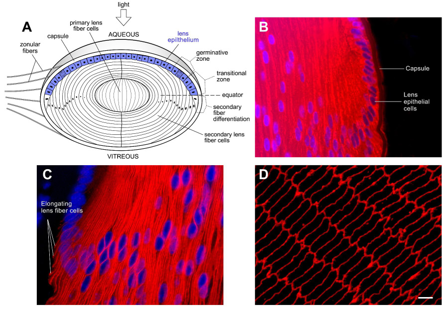
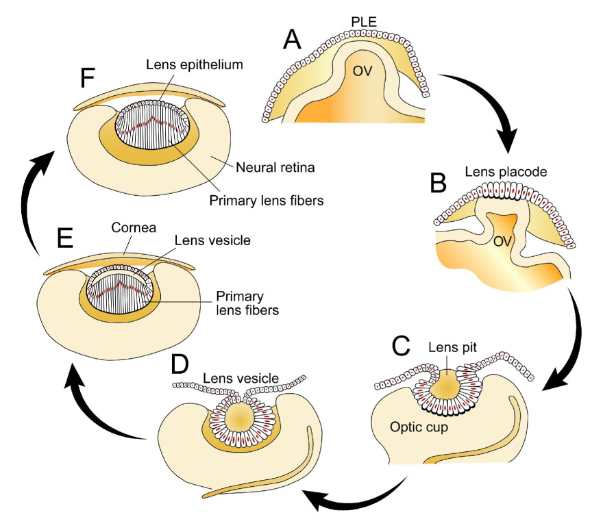
Lens epithelial cells are the parental cells responsible for growth and development of the transparent ocular lens. Many elegant investigations into their biology have focused on the factors that initiate and regulate lens epithelial cell differentiation. Because they serve key transport and cell maintenance functions throughout life, and are the primary source of metabolic activity in the lens, mechanisms to maintain lens epithelial cell integrity and survival are critical for lens transparency. The molecular chaperones alpha-crystallins are abundant proteins synthesized in the differentiated lens fiber cell cytoplasm. However, their expression in lens epithelial cells has only been appreciated very recently. Besides their important roles in the refractive and light focusing properties of the lens, alpha-crystallins have been implicated in a number of non-refractive pathways including those involving stress response, apoptosis and cell survival. The most convincing evidence for their importance in the lens epithelium has been shown by studies on the properties of lens epithelial cells from alphaA and alphaB-crystallin gene knockout mice. Novel combination of genetics, cell and molecular biology should lead to a greater understanding of how lens epithelial cells proliferate, differentiate and survive.


Figures

Figure 1
Figure 1
The lens has a unique cellular architecture consisting of a single layer of cuboidal epithelial cells which divide and differentiate into lens fiber cells. The tightly packed fiber cells have an elongated morphology and produce abundant levels of crystallins essential for transparency. (A) Schematic drawing of a lens sectioned along the optic axis to illustrate the location of the lens epithelial cells. Although all lens epithelial cells are capable of undergoing cell proliferation, in a mature lens, epithelial cell proliferation occurs mainly in a region above the lens equator called the germinative zone. Cells in the transitional zone have withdrawn from the cell cycle and are in transition to becoming secondary lens fiber cells. Formation of secondary lens fiber cells from epithelial cells occurs slightly posterior to the lens equator (horizontal dashed line). Throughout life, layers of newly formed fiber cells cover the older cells. As a result, older secondary fiber cells are located closer to the center of the lens. The primary fiber cells are formed during embryogenesis. Zonular fibers hold up the lens. (B–D) Immunofluorecence analysis of mouse lens sections with antibodies to αA-crystallin, the lens fiber cell-specific membrane intrinsic protein (MIP/AQP0) and Alexa568-labeled secondary antibodies. Nuclei were labeled with the DNA labeling fluorescent dye, DAPI. (B) αA-crystallin expression in the mid-sagittal section of a 5 day old mouse lens by immunofluorescence analysis. DNA is in blue. αA-crystallin is in red. Note that αA-crystallin immunostaining can be found in the cell cytoplasm of both lens epithelial and fiber cells. (C) In the equatorial region, the expression of lens fiber cell specific membrane protein MIP/AQP0 shown in red, marks the onset of differentiation, and elongation, of epithelial cells. (D) The bulk of the lens is composed of fiber cells which appear as hexagons in a cross-sectional slice of the lens. immunofluorescence staining for MIP is shown in red.
Figure 2
Figure 2
Embryonic development of the lens. (A) The vertebrate lens begins development as a sheet of surface ectoderm that is exposed to multiple inductive factors in the gastrulation stage of the embryo. Contact of the presumptive lens ectoderm (PLE) and the lens vesicle leads to a thickening of the PLE to form the lens placode. (B, C) Invagination of the lens placode forms the lens pit which then forms the lens vesicle. (D) Cells in the anterior of the lens vesicle proliferate and give rise to the lens epithelium while those in the posterior of the lens vesicle elongate into primary lens fiber cells. (E) Secondary fiber cells are formed by proliferation and differentiation of the lens epithelial cells near the equator. (F) The lens then grows throughout the life of a vertebrate by continued addition of lens fiber cells on top of the older fiber cells. There is no turnover in the lens cells and thus secondary fiber cells persist for life. (Adapted from Robinson, 2006; see text for further details).

References

    1. Andley UP, Song Z, Wawrousek EF, Bassnett S. The molecular chaperone alphaA-crystallin enhances lens epithelial cell growth and resistance to UVA stress. J Biol Chem. 1998;273:31252–31261. - PubMed
    1. Andley UP, Song Z, Wawrousek EF, Brady JP, Bassnett S, Fleming TP. Lens epithelial cells derived from alphaB-crystallin knockout mice demonstrate hyperproliferation and genomic instability. Faseb J. 2001;15:221–229. - PubMed
    1. Arrigo AP, Paul C, Ducasse C, Manero F, Kretz-Remy C, Virot S, Javouhey E, Mounier N, Diaz-Latoud C. Small stress proteins: novel negative modulators of apoptosis induced independently of reactive oxygen species. Prog Mol Subcell Biol. 2002;28:185–204. - PubMed
    1. Bagchi M, Katar M, Maisel H. Heat shock proteins of adult and embryonic human ocular lenses. J Cell Biochem. 2002;84:278–284. - PubMed
    1. Bai F, Xi J, Higashikubo R, Andley UP. Cell kinetic status of mouse lens epithelial cells lacking alphaA- and alphaB-crystallin. Mol Cell Biochem. 2004;265:115–122. - PubMed

Publication types

LinkOut - more resources