. 2007 Dec 26;104(52):20990-5.
doi: 10.1073/pnas.0710527105. Epub 2007 Dec 19.

Mutation of an A-kinase-anchoring protein causes long-QT syndrome

Affiliations

Mutation of an A-kinase-anchoring protein causes long-QT syndrome

Lei Chen et al. Proc Natl Acad Sci U S A. .

Abstract

A-kinase anchoring proteins (AKAPs) recruit signaling molecules and present them to downstream targets to achieve efficient spatial and temporal control of their phosphorylation state. In the heart, sympathetic nervous system (SNS) regulation of cardiac action potential duration (APD), mediated by beta-adrenergic receptor (betaAR) activation, requires assembly of AKAP9 (Yotiao) with the I(Ks) potassium channel alpha subunit (KCNQ1). KCNQ1 mutations that disrupt this complex cause type 1 long-QT syndrome (LQT1), one of the potentially lethal heritable arrhythmia syndromes. Here, we report identification of (i) regions on Yotiao critical to its binding to KCNQ1 and (ii) a single putative LQTS-causing mutation (S1570L) in AKAP9 (Yotiao) localized to the KCNQ1 binding domain in 1/50 (2%) subjects with a clinically robust phenotype for LQTS but absent in 1,320 reference alleles. The inherited S1570L mutation reduces the interaction between KCNQ1 and Yotiao, reduces the cAMP-induced phosphorylation of the channel, eliminates the functional response of the I(Ks) channel to cAMP, and prolongs the action potential in a computational model of the ventricular cardiocyte. These reconstituted cellular consequences of the inherited S1570L-Yotiao mutation are consistent with delayed repolarization of the ventricular action potential observed in the affected siblings. Thus, we have demonstrated a link between genetic perturbations in AKAP and human disease in general and AKAP9 and LQTS in particular.


Conflict of interest statement

The authors declare no conflict of interest.

Figures
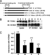
Fig. 1.
Fig. 1.
Identification of KCNQ1-binding domains on Yotiao. (A) Two key binding sites for KCNQ1 on Yotiao, one on the N terminus (residues 29–46) and the other on the C terminus (residues 1,574–1,643), are shown in a schematic diagram. Four Yotiao constructs were used in the IP experiments to study the Yotiao/KCNQ1 interaction as shown in the diagram and described in Results. (B) A representative IP experiment demonstrates that both Yotiao N and C termini participate in the interaction with KCNQ1. An anti-KCNQ1 antibody was used to precipitate the KCNQ1/Yotiao complex. Control experiment was performed by using nonspecific goat IgG. Western blots (IB) of the lysates are shown (Left). Western blots of the immunocomplex are shown (Right). (C) The interactions between KCNQ1 and WT or mutant Yotiao were quantified by measuring Western blots of the immunocomplex, after correcting the IP input errors. Results were all normalized to WT Yotiao. *, P < 0.001; ANOVA and Bonferroni test.
Fig. 2.
Fig. 2.
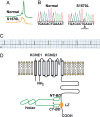
A missense variant, S1570L, in AKAP9-encoded Yotiao in human LQTS. (A) DHPLC analysis of the DNAs from the patient positive for S1570L-Yotiao and normal controls. (B) Chromatograms of DNA sequences of the patient positive for S1570L-Yotiao and normal control. Underlined G-to-A change in nucleotide sequence causes the S1570L (serine, S, to leucine, L) missense mutation in Yotiao. (C) ECG of S1570L-Yotiao-positive patient with symptomatic LQTS (QTc, 485 ms). (D) A schematic diagram of the IKs/Yotiao complex. Shown in gray color in the plasma membrane are KCNQ1 and KCNE1, α- and β-subunits of IKs, respectively. A leucine zipper motif (LZ) is located at the C terminus of KCNQ1 and is the binding site for Yotiao. AKAP Yotiao is depicted in green. NT-BD and CT-BD indicate the two KCNQ1-binding sites on Yotiao N and C termini. The LQTS-associated mutation S1570L is located close to the CT-BD, indicated by an arrow.
Fig. 3.
Fig. 3.
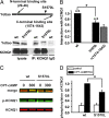
S1570L Yotiao reduces the interaction with and the cAMP-dependent phosphorylation of KCNQ1. (A) An anti-KCNQ1 antibody was used to precipitate the KCNQ1/Yotiao complex from the lysates of the transfected CHO cells. A control experiment was performed by using nonspecific goat IgG. Western blots (IB) of the lysates are shown (Left). Western blots of the immunocomplex are shown (Right). (B) The interactions between KCNQ1 and either WT- or S1570L-Yotiao are quantified by measuring Western blots of the immunocomplex after correcting the IP input errors. Results are normalized to WT-Yotiao. The S1570L mutation significantly reduced the interaction between KCNQ1. *, P < 0.05 (paired t test). Yotiao and KCNQ1. *, P < 0.05 (paired t test). The gray bar represents the interaction between KCNQ1 and Yotiao Δ(1,574–1,643) reproduced from Fig. 1 and is for comparison purposes only. (C) Phosphorylation of KCNQ1 was assayed in the cells expressing KCNQ1 with either WT- or S1570L-Yotiao. Lysates were analyzed by dual Western blot by using an infrared imaging system. Green signals (Middle) detect phosphorylated KCNQ1 (p-KCNQ1) protein. Red signals (Bottom) detect total KCNQ1 protein. A merged view is presented (Top). (D) S27 phosphorylated KCNQ1 is quantified by measuring the relative band intensity on the phospho-KCNQ1 Western blots after correcting for total KCNQ1 loading. *, P < 0.01.
Fig. 4.
Fig. 4.
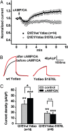
S1570L-Yotiao markedly inhibits the functional response of IKs channels to cAMP. (A) Perforated-patch recordings were performed on CHO cells transiently expressing KCNQ1 and KCNE1 with either WT- (filled squares) or S1570L-Yotiao (open circles). Application of CPT-cAMP/OA was indicated by the arrow. Tail current amplitude was monitored and normalized to the current amplitude before application of CPT-cAMP/OA. Normalized tail current amplitude is plotted against time. (B) Average IKs current traces before (black) and after (red) application of CPT-cAMP/OA in cells transfected with WT-Yotiao (Left) or S1570L-Yotiao (Right) were shown. (C) Comparison of IKs tail current densities before (control, gray bar) and after (black bar) CPT-cAMP/OA treatment in cells transfected with either WT- or S1570L-Yotiao. Cells transfected with WT-Yotiao showed an increase in current density after CPT-cAMP/OA treatment (*, P < 0.01; control vs. cAMP/OA, paired t test). Cells transfected with S1570L-Yotiao did not respond to cAMP/OA (n.s. indicates no significant difference; control vs. cAMP/OA, paired t test). Basal (control) and cAMP/OA-treated IKs current densities in cells transfected with S1570L-Yotiao are significantly lower than that of the cells transfected with WT-Yotiao (**, P < 0.05; WT control vs. S1570L control, Welch's t test; ***, P < 0.01; WT cAMP/OA vs. S1570L cAMP/OA, Welch's t test).
Fig. 5.
Fig. 5.
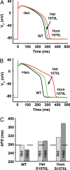
S1570L-Yotiao is predicted to prolong action potential duration. (A) Simulations for steady-state action potentials stimulated at 1 Hz in WT (black line), heterozygous S1570L (green line), and homozygous S1570L (red line) cells show that the loss of functional translation of basal IKs complex phosphorylation leads to a APD prolongation (+35 ms for the homozygous cell). (B) In the presence of maximal isoproterenol stimulation, APD prolongation is severe in the mutant cells (+121 ms for the homozygous cell). (C) Significant APD prolongation is predicted in a gene dosage- and adrenergic stimulation-dependant manner.

Similar articles

Cited by

References

    1. McConnachie G, Langeberg LK, Scott JD. Trends Mol Med. 2006;12:317–323. - PubMed
    1. Dodge-Kafka KL, Langeberg L, Scott JD. Circ Res. 2006;98:993–1001. - PubMed
    1. Smith FD, Langeberg LK, Scott JD. Trends Biochem Sci. 2006;31:316–323. - PubMed
    1. Marx SO, Reiken S, Hisamatsu Y, Jayaraman T, Burkhoff D, Rosemblit N, Marks AR. Cell. 2000;101:365–376. - PubMed
    1. Hulme JT, Westenbroek RE, Scheuer T, Catterall WA. Proc Natl Acad Sci USA. 2006;103:16574–16579. - PMC - PubMed

Publication types

MeSH terms