

Three-dimensional structure of cytoplasmic dynein bound to microtubules
- PMID: 18093913
- PMCID: PMC2409227
- DOI: 10.1073/pnas.0710406105
Three-dimensional structure of cytoplasmic dynein bound to microtubules
Abstract
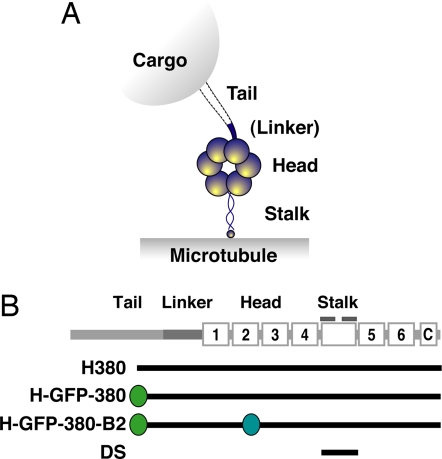
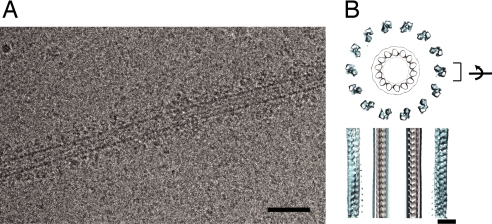
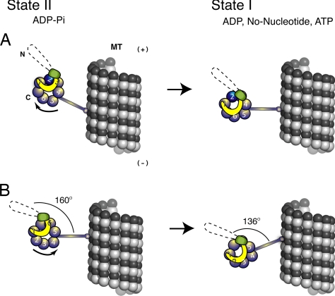
Cytoplasmic dynein is a large, microtubule-dependent molecular motor (1.2 MDa). Although the structure of dynein by itself has been characterized, its conformation in complex with microtubules is still unknown. Here, we used cryoelectron microscopy (cryo-EM) to visualize the interaction between dynein and microtubules. Most dynein molecules in the nucleotide-free state are bound to the microtubule in a defined conformation and orientation. A 3D image reconstruction revealed that dynein's head domain, formed by a ring-like arrangement of AAA+ domains, is located approximately 280 A away from the center of the microtubule. The order of the AAA+ domains in the ring was determined by using recombinant markers. Furthermore, a 3D helical image reconstruction of microtubules with a dynein's microtubule binding domain [dynein stalk (DS)] revealed that the stalk extends perpendicular to the microtubule. By combining the 3D maps of the dynein-microtubule and DS-microtubule complexes, we present a model for how dynein in the nucleotide-free state binds to microtubules and discuss models for dynein's power stroke.
Conflict of interest statement
The authors declare no conflict of interest.
Figures


Similar articles
-
Dynein and kinesin share an overlapping microtubule-binding site.EMBO J. 2004 Jul 7;23(13):2459-67. doi: 10.1038/sj.emboj.7600240. Epub 2004 Jun 3. EMBO J. 2004. PMID: 15175652 Free PMC article.
-
Distinct functions of nucleotide-binding/hydrolysis sites in the four AAA modules of cytoplasmic dynein.Biochemistry. 2004 Sep 7;43(35):11266-74. doi: 10.1021/bi048985a. Biochemistry. 2004. PMID: 15366936
-
Structural basis for microtubule binding and release by dynein.Science. 2012 Sep 21;337(6101):1532-1536. doi: 10.1126/science.1224151. Science. 2012. PMID: 22997337 Free PMC article.
-
Communication between the AAA+ ring and microtubule-binding domain of dynein.Biochem Cell Biol. 2010 Feb;88(1):15-21. doi: 10.1139/o09-127. Biochem Cell Biol. 2010. PMID: 20130675 Free PMC article. Review.
-
The role of the dynein stalk in cytoplasmic and flagellar motility.Eur Biophys J. 1998;27(5):466-73. doi: 10.1007/s002490050157. Eur Biophys J. 1998. PMID: 9760728 Review.
Cited by
-
Overview of the mechanism of cytoskeletal motors based on structure.Biophys Rev. 2018 Apr;10(2):571-581. doi: 10.1007/s12551-017-0368-1. Epub 2017 Dec 12. Biophys Rev. 2018. PMID: 29235081 Free PMC article. Review.
-
The Ndc80 kinetochore complex forms oligomeric arrays along microtubules.Nature. 2010 Oct 14;467(7317):805-10. doi: 10.1038/nature09423. Nature. 2010. PMID: 20944740 Free PMC article.
-
The N-terminal coiled-coil of Ndel1 is a regulated scaffold that recruits LIS1 to dynein.J Cell Biol. 2011 Feb 7;192(3):433-45. doi: 10.1083/jcb.201011142. Epub 2011 Jan 31. J Cell Biol. 2011. PMID: 21282465 Free PMC article.
-
Analyses of dynein heavy chain mutations reveal complex interactions between dynein motor domains and cellular dynein functions.Genetics. 2012 Aug;191(4):1157-79. doi: 10.1534/genetics.112.141580. Epub 2012 May 29. Genetics. 2012. PMID: 22649085 Free PMC article.
-
A single protofilament is sufficient to support unidirectional walking of dynein and kinesin.PLoS One. 2012;7(8):e42990. doi: 10.1371/journal.pone.0042990. Epub 2012 Aug 10. PLoS One. 2012. PMID: 22900078 Free PMC article.
References
-
- Mallik R, Carter BC, Lex SA, King SJ, Gross SP. Cytoplasmic dynein functions as a gear in response to load. Nature. 2004;427:649–652. - PubMed
-
- Vallee RB, Williams JC, Varma D, Barnhart LE. Dynein: An ancient motor protein involved in multiple modes of transport. J Neurobiol. 2004;58:189–200. - PubMed
-
- Kon T, Mogami T, Ohkura R, Nishiura M, Sutoh K. ATP hydrolysis cycle-dependent tail motions in cytoplasmic dynein. Nat Struct Mol Biol. 2005;12:513–519. - PubMed
Publication types
MeSH terms
Substances
Grants and funding
LinkOut - more resources
Full Text Sources
Other Literature Sources
