. 2008 Jan 8;105(1):13-9.
doi: 10.1073/pnas.0710504105. Epub 2007 Dec 18.

EPLIN mediates linkage of the cadherin catenin complex to F-actin and stabilizes the circumferential actin belt

Affiliations

EPLIN mediates linkage of the cadherin catenin complex to F-actin and stabilizes the circumferential actin belt

Kentaro Abe et al. Proc Natl Acad Sci U S A. .

Abstract

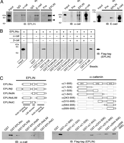
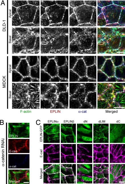
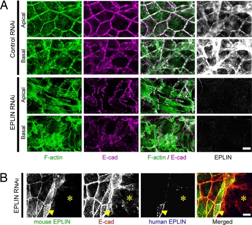
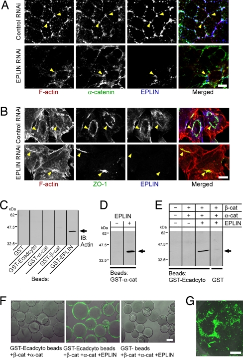
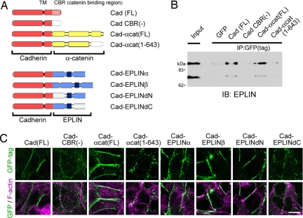
The cadherin-catenin complex is the major machinery for cell-cell adhesion in many animal species. This complex in general associates with actin fibers at its cytoplasmic side, organizing the adherens junction (AJ). In epithelial cells, the AJ encircles the cells near their apical surface and forms the "zonula adherens" or "adhesion belt." The mechanism as to how the cadherin-catenin complex and F-actin cooperate to generate these junctional structures, however, remains unknown. Here, we show that EPLIN (epithelial protein lost in neoplasm; also known as Lima-1), an actin-binding protein, couples with alpha-catenin and, in turn, links the cadherin-catenin complex to F-actin. Without EPLIN, this linkage was unable to form. When EPLIN had been depleted in epithelial cells, the adhesion belt was disorganized and converted into zipper-like junctions in which actin fibers were radially arranged. However, nonjunctional actin fibers were not particularly affected by EPLIN depletion. As EPLIN is known to have the ability to suppress actin depolymerization, our results suggest that EPLIN functions to link the cadherin-catenin complex to F-actin and simultaneously stabilizes this population of actin fibers, resulting in the establishment of the adhesion belt.


Conflict of interest statement

The authors declare no conflict of interest.

Figures

Fig. 1.
Fig. 1.
EPLIN interacts with α-catenin to couple to the cadherin–β-catenin complex. (A) Lysates of DLD-1 cells were subjected to immunoprecipitation (IP) with antibodies against α-catenin (α-cat), E-cadherin (E-cad), or EPLIN. As a control, preimmune serum (Pre), or mouse or rabbit anti-GFP IgGs (IgG) were used. The precipitates were analyzed by SDS/PAGE and Western blotting to detect α-cat, E-cad, or EPLIN. Arrowheads indicate the α (Lower) and β (Upper) isoforms of EPLIN. In Input, 2% of the lysate used for immunoprecipitation was loaded in each experiment. IB, immunoblot. (B) In vitro binding between purified proteins. Recombinant EPLINα-Flag proteins, from which the GST tag had been removed (SI Fig. 7), were incubated with various GST-fusion protein-coated beads, as indicated. The arrow points to the pulled-down EPLIN, detected by Western blotting with antibodies against Flag tag. EPLIN efficiently coprecipitates with GST-α-cat, but not with GST, GST-β-cat (β-catenin), or GST-Ecadcyto. In the presence of both α-catenin and β-catenin (GST tag removed), however, EPLIN can be pulled down by GST-Ecadcyto. (C) In vitro binding between purified fragments of α-catenin and EPLINα-Flag, carrying deletions of each protein. The diagrams show the deletion series of EPLIN (Left) and α-catenin (Right) (see also SI Fig. 7). A GST tag was fused to the N terminus of each molecule. The interaction of the proteins was analyzed by Western blotting with antibodies against Flag tag (for EPLIN) or α-catenin. All constructs having the VH3 domain bound EPLIN. A faint interaction of α (amino acids 1–643) with EPLIN was detectable, but it was not reproducible.
Fig. 2.
Fig. 2.
EPLIN localization at cell–cell junctions requires α-catenin. (A) Distribution of EPLIN, α-catenin, and F-actin, which are triple-stained, in DLD-1 or MDCK cells. Confocal sections focused around the apical-most and basal-most portions of the cells are shown. These three proteins colocalize well in the apical level, but not in the basal level, of the cells. In particular, EPLIN is not detectable along actin stress fibers in MDCK cells. The distributions of E-cadherin and α-catenin are essentially identical; therefore, only the data for either one of them are shown. (B) DLD-1 cells transfected with siRNA against α-catenin do not show the colocalization of E-cadherin and EPLIN. Depletion of α-catenin was confirmed by Western blot analysis (SI Fig. 8). (C) DLD-1 cells stably transfected with various EPLIN constructs shown in Fig. 1C. EPLINα, EPLINβ, and EPLINdLIM show cell junctional accumulation, but EPLINdN and EPLINdC do not. (Scale bars: 10 μm.)
Fig. 3.
Fig. 3.
EPLIN is indispensable for adhesion belt formation. (A) EPLIN depletion disrupts the apical organization of F-actin. DLD-1 cells transfected with control siRNA or siRNA against EPLIN were triple-stained for F-actin, E-cadherin, and EPLIN. Apical-most and basal-most confocal sections are shown. Note that the circular arrangement of F-actin along the adhesion belt is converted into a radial one by EPLIN depletion, which was confirmed by Western blot analysis (SI Fig. 7). (B) Rescue of the EPLIN-depletion phenotypes by mouse EPLIN expression. DLD-1 cells were transfected with mouse EPLINα-GFP cDNA; and then, a mixed culture of cells expressing and not expressing mouse EPLIN was treated with siRNA against human EPLIN. Green, mouse EPLIN; magenta, E-cadherin; blue, human EPLIN. Asterisks indicate a portion of the culture not expressing mouse EPLIN. In this portion, E-cadherin distribution is disorganized; whereas, in the mouse EPLIN-positive area, E-cadherin remains to organize the adhesion belts. The arrow points to a cell that escaped from the siRNA treatment, resulting in expression of both mouse and human EPLIN. The EPLIN antibodies used here recognize only the human EPLIN. (Scale bars: 10 μm.)
Fig. 4.
Fig. 4.
EPLIN stabilizes apical actin bundles and links them to the cadherin–catenin complex. (A) EPLIN depletion enhances the latrunculin A sensitivity of F-actin at AJ. DLD-1 cells treated with control or EPLIN siRNA were incubated with 1 μM latrunculin A for 30 min, then fixed, and triple-stained for F-actin, α-catenin, and EPLIN. F-actin remains as clusters in the control cultures, colocalizing with α-catenin and EPLIN, as indicated by the arrowheads. When EPLIN is depleted, F-actin and α-catenin becomes dispersed. EPLIN expression is not down-regulated in some cells in the EPLIN RNAi cultures, and these cells maintain actin clusters (arrowheads). (B) Apical actin bundles in α-catenin-deficient cells are sensitive to EPLIN depletion. R2/7 cells were treated with control or EPLIN siRNA, fixed, and triple-stained for F-actin, ZO-1, and EPLIN. Arrowheads in the control show examples of ZO-1-positive F-actin bundles, where EPLIN is also localized. In EPLIN-depleted cells, the ZO-1 positive structures are collapsed, as indicated by the arrowheads. (C–E) In vitro binding between F-actin and various proteins. GST-fusion protein-coated beads were incubated with 4 μg of polymerized actin (5–10 μm long), and collected by centrifugation at 3,500 × g. The precipitates were washed extensively and subjected to Western blot assays for detecting the bound actin (arrows). In D, GST-α-catenin-coated beads were preincubated with purified EPLIN, washed, and then used for the above assay. In E, GST-Ecadcyto-coated beads were sequentially incubated with recombinant β-catenin, α-catenin, and EPLIN, before use for the F-actin pull-down assay. Ecadcyto can interact with actin, but only when β-catenin, α-catenin, and EPLIN are present all together. (F and G) Beads incubated with F-actin were stained with Alexa Fluor-488-conjugated phalloidin, followed by observations with a confocal fluorescence microscope. In G, a higher magnification view of the surface of a GST-Ecadcyto bead incubated with β-catenin, α-catenin, and EPLIN is shown, on which filamentous actins are deposited. (Scale bars: A, B, and G, 10 μm; F, 50 μm.)
Fig. 5.
Fig. 5.
The cadherin–EPLIN association is sufficient to link junctional actin rings to cadherin. (A) Diagrams showing fusion constructs between cadherin and α-catenin (Upper) (13) or EPLIN (Lower). To use these constructs for generating transfectants, a GFP tag was fused to the C terminus of each molecule. (B) Binding of EPLIN to cadherin-α-catenin fusion proteins. L cells were stably transfected with cadherin or the cadherin-α-catenin fusion proteins. These molecules were immunoprecipitated with antibodies against the GFP tag from a lysate of each transfectant, and the precipitates were analyzed to detect EPLIN by Western blotting. (C) L cells stably expressing GFP-tagged cadherin (Upper) or cadherin fusion proteins (Lower) indicated in A were double-stained for the GFP tag and F-actin. (Scale bar: 10 μm.)

References

    1. Mege RM, Gavard J, Lambert M. Curr Opin Cell Biol. 2006;18:541–548. - PubMed
    1. Lecuit T. Trends Cell Biol. 2005;15:34–42. - PubMed
    1. Pokutta S, Weis WI. Annu Rev Cell Dev Biol. 2007;23:237–261. - PubMed
    1. Hildebrand JD. J Cell Sci. 2005;118:5191–5203. - PubMed
    1. Haigo SL, Hildebrand JD, Harland RM, Wallingford JB. Curr Biol. 2003;13:2125–2137. - PubMed

Publication types