. 2007 Dec 26;104(52):20973-8.
doi: 10.1073/pnas.0710316105. Epub 2007 Dec 18.

Nck adaptor proteins control the organization of neuronal circuits important for walking

Affiliations

Nck adaptor proteins control the organization of neuronal circuits important for walking

James P Fawcett et al. Proc Natl Acad Sci U S A. .

Abstract

The intracellular signaling targets used by mammalian axon guidance receptors to organize the nervous system in vivo are unclear. The Nck1 and Nck2 SH2/SH3 adaptors (collectively Nck) can couple phosphotyrosine (pTyr) signals to reorganization of the actin cytoskeleton and are therefore candidates for linking guidance cues to the regulatory machinery of the cytoskeleton. We find that selective inactivation of Nck in the murine nervous system causes a hopping gait and a defect in the spinal central pattern generator, which is characterized by synchronous firing of bilateral ventral motor neurons. Nck-deficient mice also show abnormal projections of corticospinal tract axons and defective development of the posterior tract of the anterior commissure. These phenotypes are consistent with a role for Nck in signaling initiated by different classes of guidance receptors, including the EphA4 receptor tyrosine kinase. Our data indicate that Nck adaptors couple pTyr guidance signals to cytoskeletal events required for the ipsilateral projections of spinal cord neurons and thus for normal limb movement.


Conflict of interest statement

The authors declare no conflict of interest.

Figures
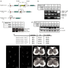
Fig. 1.
Fig. 1.
Generation and characterization of mice containing a conditional allele of Nck2. (a) Schematic diagram of the strategy used to generate the conditional mutant of Nck2. (b) PCR genotyping of mice generated by crossing a Nck1−/−;Nck2flx/flx female with a Nck1+/−;Nck2flx/flx;Nestin-Cre male. An Nck1+/−;Nck2flx/wt;Nestin-Cre animal was included as a control (last lane). Top gel, Nck1 allele; middle gel, Nck2 allele; bottom gel, Cre. (c) Proteins isolated from the brain (top gel) or liver (bottom gel) of individual P1 mice, as noted, were immunoblotted with a pan-Nck antibody. (d) Dissociated cortical neurons from embryonic day-16 animals of various genotypes, as noted, were immunoblotted with a pan-Nck antibody (lower band, upper gel) and reprobed with tubulin (lower gel). Note the loss of Nck expression with decreasing Nck alleles [lanes 1–3 (0 alleles) < lane 7 (2 alleles) < lanes 4–6 (3 alleles)]. (e) Mendelian ratio of the offspring generated from crossing Nck1−/−;Nck2flx/flx females with Nck1+/−;Nck2flx/flx;Nestin-Cre males (n > 100 animals at each time point). (f and g) Hindpaw prints from a 12-week-old control mouse (Nck1−/−;Nck2flx/flx) (f) or a Nck-deficient mouse (Nck1−/−;Nck2flx/flx;Nestin-Cre) (g). (h–k) Dark-field photographs of spinal cord cross-sections at the thoracic (h and i) and lumbar (j and k) level of a control (Nck1−/−;Nck2flxflx) (h and j) and Nck-deficient (i and k) mouse. cc, central canal; DGM, dorsal gray matter.
Fig. 2.
Fig. 2.
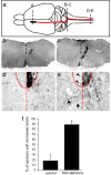
Reduction of Nck in the developing CNS leads to aberrant axon migration of corticospinal tract fibers. (a) Schematic diagram depicting sites of tracer injection in the somatomotor cortex (A) and path of the CST axons from the cortex across the medullary decussation (B-C) into the spinal cord (D-E). (b and c) BDA-labeled axons are found at the spinomedullary junction (b), CST axons decussate in the pyramidal decussation (c). (d and e) Unilateral labeling of CST-positive fibers in the ventral aspect of the DF in both control (Nck1−/−;Nck2flx/flx) (d) and Nck-deficient (e) animals at the thoracic level. (e) In the Nck-deficient mice, a number of fibers are seen to recross the midline in the gray matter (arrows); in addition, a few fibers were seen crossing in the white matter (arrowheads). The dashed line denotes the spinal midline, and the solid line denotes DF. (f) Quantification of recrossing fibers in control (Nck1−/−;Nck2flx/flx) and Nck-deficient mice (n = 3 for each genotype; 70–120 slices per animal). Error bars indicate SD; *, P < 0.01 compared with control (Student's t test).
Fig. 3.
Fig. 3.
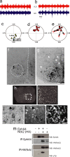
Loss of Nck leads to synchronous firing in bilateral ventral roots and defects in commissural axon guidance. (a and b) Representative trace recordings from the left (lL5) and right (rL5) ventral roots at the L5 level after the addition of NMDA and serotonin to isolated spinal cords from P2 control (Nck1+/−;Nck2flx/flx;Nestin-Cre) (a) and an Nck-deficient (Nck1−/−;Nck2flx/flx;Nestin-Cre) (b) mouse. (c and d) Circular-phase diagrams from control [Nck1+/−;Nck2flx/flx;Nestin-Cre (yellow triangles; n = 5); Nck1−/−;Nck2flx/flx (green triangle; n = 1); or Nck1+/−;Nck2flx/flx (blue triangle; n = 1)] (c) and Nck-deficient [Nck1−/−;Nck2flx/flx;Nestin-Cre (red circles; n = 8)] (d) mice. (e) Circular-phase diagram of Nck-deficient mice after the addition of 100 μM sarcosine. Filled red circles represent averaged vector plots of individual animals before sarcosine treatment. Open circles identify the final vector plot of individual animals after 20 min of sarcosine treatment. The arrows mark the movement of an individual animal's vector profiles after treatment. All recordings shown are from the L5 level. (f and g) Epifluorescent images of coronal sections through the L2 spinal cord after contralateral caudal application of rhodamine dextran amine (molecular weight, 3,000) crystals at the L4 level in control Nck1−/−;Nck2flx/flx (f) and Nck-deficient (g) mice. Descending CINs are found within the dashed circles. Dashed lines mark the midline. Arrows in g indicate fibers crossing the midline in Nck-deficient animals. (h and i) Nck1-β-gal expression of coronal sections through the lumbar spinal cord of an adult mouse. (i) Enlargement of boxed region shown in h. (j and k) EphA4-positive cells in the medial–ventrolateral spinal cord (j, arrows) that are Nck1-positive (k, arrows) as revealed by EphA4 RNA in situ hybridization (j) and β-gal antibodies (k). (l) Hoechst staining to show nuclei. (Scale bar: 10 μm.) Not all Nck1-positive cells are EphA4-positive, (j and k, asterisks), nor are all EphA4- positive cells Nck1-positive (j and k, arrowheads). (m) HEK293T cells were transfected as indicated. Lysates were immunoprecipitated (IP) with antibodies as indicated and probed with anti-EphA4 (top row), anti-HA antibodies (middle two rows), or anti-pTyr antibodies (bottom row).
Fig. 4.
Fig. 4.
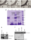
Nck proteins have multiple functions and interactions in the nervous system. (a–c) Loss of Nck leads to defective development of the posterior tract of the anterior commissure. Shown are bright-field images of horizontal sections through the anterior commissure (AC) of 12-week-old animals. Anterior is up. Anterior (aAC), posterior (pAC, indicated by arrows), and a third smaller tract ventral to the pAC fibers (arrowheads) are indicated. (d) Colloidal Coomassie stained gel after GST fusion pull-downs as outlined. Lane 1, GST-Nck1-SH3 alone; lane 2, GST alone; lane 3, GST-Nck1-SH3 GST and mouse brain lysate; lane 4, GST and mouse brain lysate. Proteins identified by liquid chromatography tandem MS (LC-MS/MS) are shown beside each arrow. (e) Rat brain lysate was immunoprecipitated as indicated. Proteins were detected by immunoblotting with α-chimaerin antibodies (Upper) or Nck antibodies (Lower). (f) Brain lysate was mixed with GST or recombinant GST fusion proteins as indicated. (Upper) The immunoblot was probed for α-chimaerin. (Lower) Ponceau S stain of blot to show level of fusion protein.

Similar articles

Cited by

References

    1. Jones N, Blasutig IM, Eremina V, Ruston JM, Bladt F, Li H, Huang H, Larose L, Li SS, Takano T, et al. Nature. 2006;440:818–823. - PubMed
    1. Li W, Fan J, Woodley DT. Oncogene. 2001;20:6403–6417. - PubMed
    1. Holland SJ, Gale NW, Gish GD, Roth RA, Songyang Z, Cantley LC, Henkemeyer M, Yancopoulos GD, Pawson T. EMBO J. 1997;16:3877–3888. - PMC - PubMed
    1. Stein E, Huynh-Do U, Lane AA, Cerretti DP, Daniel TO. J Biol Chem. 1998;273:1303–1308. - PubMed
    1. Cowan CA, Henkemeyer M. Nature. 2001;413:174–179. - PubMed

Publication types

LinkOut - more resources