. 2007 Dec 26;104(52):20902-7.
doi: 10.1073/pnas.0708541104. Epub 2007 Dec 18.

Defects in XRCC4 and KU80 differentially affect the joining of distal nonhomologous ends

Affiliations

Defects in XRCC4 and KU80 differentially affect the joining of distal nonhomologous ends

Josée Guirouilh-Barbat et al. Proc Natl Acad Sci U S A. .

Abstract

XRCC4-null mice have a more severe phenotype than KU80-null mice. Here, we address whether this difference in phenotype is connected to nonhomologous end-joining (NHEJ). We used intrachromosomal substrates to monitor NHEJ of two distal double-strand breaks (DSBs) targeted by I-SceI, in living cells. In xrcc4-defective XR-1 cells, a residual but significant end-joining process exists, which primarily uses microhomologies distal from the DSB. However, NHEJ efficiency was strongly reduced in xrcc4-defective XR-1 cells versus complemented cells, contrasting with KU-deficient xrs6 cells, which showed levels of end-joining similar to those of complemented cells. Nevertheless, sequence analysis of the repair junctions indicated that the accuracy of end-joining was strongly affected in both xrcc4-deficient and KU-deficient cells. More specifically, these data showed that the KU80/XRCC4 pathway is conservative and not intrinsically error-prone but can accommodate non-fully complementary ends at the cost of limited mutagenesis.


Conflict of interest statement

The authors declare no conflict of interest.

Figures

Fig. 1.
Fig. 1.
The NHEJ substrate. (A) The reporter genes are H2Kd and CD4, coding for membrane antigens. Before expression of the meganuclease I-SceI, only H2Kd is expressed, because CD4 is too far from the pCMV promoter. I-SceI cleaves the two sites on either side of an internal fragment containing H2Kd. The deletion of this fragment and the joining of the two distal ends places the CD4 gene directly downstream of the promoter, and CD4 is then expressed. The expression of the H2Kd and CD4 antigens can be monitored by FACS or by immunofluorescence. Cells having undergone NHEJ events can be selected by magnetic cell sorting, using antibodies against CD4; the repaired junctions can be amplified by PCR and sequenced. Importantly, the two colinear I-SceI cleavage sites of the NHEJ substrate are located in noncoding sequences, and the events are not selected for their viability. Therefore, this strategy allows analysis of the accuracy of joining of distal ends (1). (B) Two substrates were constructed. In pCOH-CD4, the two I-SceI sites are in direct orientation, and I-SceI cleavage generates fully complementary ends for CD4 expression (Left). End-joining either is accurate or leads to extended deletion (sometimes associated with DNA capture). In pINV-CD4, the I-SceI sites are in inverted orientation and generate non-fully complementary ends for CD4 expression (Right). DSB repair either leads to extended deletion or uses the annealing of 2 of the 4 protruding nucleotides (in blue with blue dot), according to 3 classes of intermediates: class I contains 1 nucleotide gap (green -) and one mismatch (red A/A); class II contains two A/A mismatches (in red) and two nicks (black triangles); and class III contains two 3′ nonannealed tails (in red). (C) Names and descriptions of the cell lines used.
Fig. 2.
Fig. 2.
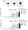
Frequency of NHEJ. (A) Example of FACS analysis of I-SceI-induced NHEJ in xrcc4-defective cells in absence of I-SceI (Left), after I-SceI transfection but in absence of complementation by XRCC4 (Center); and after both I-SceI transfection and XRCC4 complementation (Right). (B) Excision/deletion (CD4+) involving fully complementary ends. (C) Excision/deletion (CD4+) involving non-fully complementary ends. The values correspond to at least five experiments. *, Significant statistical difference (P < 0.05) between the control (without I-SceI) and induced by I-SceI. **, Significant statistical difference (P < 0.05) by complementation with XRCC4. Error bars depict the SEM. The names of the clones used are indicated in the figure.
Fig. 3.
Fig. 3.
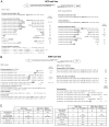
Sequence analysis of the junctions in xrcc4-defective cells. (A) Junction sequences from fully complementary ends, in xrcc4-defective cells. (B) Junction sequences from non-fully complementary ends, xrcc4-defective cells. Sequences were performed by using at least two independent sets of experiments. The I-SceI sites are in bold. Squares indicate the locations of internal microhomologies. The numbers of nucleotides involved in microhomology annealing are indicated on the right part of each sequence. Parentheses indicate the number of identical sequences. (C) Frequencies of the different events. The values between the parentheses correspond to the percentage of deletion, using microhomologies among the deletion events.
Fig. 4.
Fig. 4.
Junction sequences of non-fully complementary ends in KU80-defective cells (xrs6). (A) The structures of the DNA ends involved are shown at Top. In bold is the I-SceI cleavage site. Underlined are protruding nucleotides after I-SceI cleavage. Squares indicate the locations of internal microhomologies. Sequences were performed by using at least two independent sets of experiments. The I-SceI sites are in bold. The numbers of nucleotides involved in microhomology anneling are indicated on the right part of each sequence. Parentheses indicates the number of identical sequences. (B) Frequencies of the different events. The values between the parentheses correspond to the percentage of deletion, using microhomologies among the deletion events.
Fig. 5.
Fig. 5.
The canonical and NHEJ-alt pathways. (A) The canonical NHEJ pathway, involving KU and XRCC4, can seal double-strand ends, even distal and non-fully complementary ends, in a conservative fashion. (B) In the KU-alt pathway, the main event is extended deletion at the junction, generally associated with the use of internal microhomologies distant from the ends. This supports the hypothesis that the NHEJ-alt pathway is initiated by single-strand resection (or by a helicase generating single-strand tails), followed by the joining and annealing of internal sequences. The maturation of the intermediate structures should reseal the DNA, leading to extended deletions at the junction.

Comment in

  • Alternative endings.
    Haber JE. Haber JE. Proc Natl Acad Sci U S A. 2008 Jan 15;105(2):405-6. doi: 10.1073/pnas.0711334105. Epub 2008 Jan 7. Proc Natl Acad Sci U S A. 2008. PMID: 18180452 Free PMC article. No abstract available.

References

    1. Guirouilh-Barbat J, Huck S, Bertrand P, Pirzio L, Desmaze C, Sabatier L, Lopez BS. Mol Cell. 2004;14:611–623. - PubMed
    1. Lees-Miller SP, Meek K. Biochimie. 2003;85:1161–1173. - PubMed
    1. Revy P, Malivert L, de Villartay JP. Curr Opin Allergy Clin Immunol. 2006;6:416–420. - PubMed
    1. Feldmann E, Schmiemann V, Goedecke W, Reichenberger S, Pfeiffer P. Nucleic Acids Res. 2000;28:2585–2596. - PMC - PubMed
    1. Kabotyanski EB, Gomelsky L, Han JO, Stamato TD, Roth DB. Nucleic Acids Res. 1998;26:5333–5342. - PMC - PubMed

Publication types