

Effects of immunosuppressive treatment on microsomal prostaglandin E synthase 1 and cyclooxygenases expression in muscle tissue of patients with polymyositis or dermatomyositis
- PMID: 18094001
- PMCID: PMC2582339
- DOI: 10.1136/ard.2007.079525
Effects of immunosuppressive treatment on microsomal prostaglandin E synthase 1 and cyclooxygenases expression in muscle tissue of patients with polymyositis or dermatomyositis
Abstract
Objectives: To investigate the expression of microsomal prostaglandin E (PGE) synthase 1 (mPGES-1) and cyclooxygenase (COX) in muscle biopsies from patients with polymyositis or dermatomyositis before and after conventional immunosuppressive treatment.
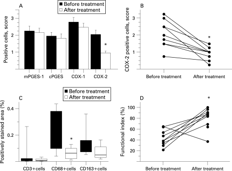
Methods: mPGES-1 and COX expression was evaluated by immunohistochemistry in muscle tissue from healthy individuals and from patients with polymyositis or dermatomyositis before and after conventional immunosuppressive treatment. The number of inflammatory cell infiltrates, T lymphocytes and macrophages was estimated before and after treatment. To localise the mPGES-1 expression double immunofluorescence was performed with antibodies against mPGES-1, CD3, CD68, CD163 and a fibroblast marker. A functional index was used to assess muscle function.
Results: In patients with myositis, mPGES-1, COX-2 and COX-1 expression was significantly higher compared to healthy individuals and associated with inflammatory cells. Double immunofluorescence demonstrated a predominant expression of mPGES-1 in macrophages. Conventional immunosuppressive treatment resulted in improved but still lower muscle function than normal. A decreased number of CD68-positive macrophages and reduced COX-2 expression in muscle tissue was also seen. By contrast, following the same treatment no significant changes were observed in muscle tissue regarding number of infiltrates, T lymphocytes, CD163-positive macrophages or mPGES-1 protein levels.
Conclusions: Increased expression of mPGES-1, COX-1 and COX-2 at protein level was observed in muscle tissue from patients with myositis compared to healthy individuals. Conventional immunosuppressive treatment led to a significant downregulation of COX-2 in myositis muscle tissue. However, the expression of mPGES-1 and COX-1 remained unchanged indicating a role of these enzymes in the chronicity of these diseases.
Conflict of interest statement
Figures


References
-
- Mastaglia FL, Garlepp MJ, Phillips BA, Zilko PJ. Inflammatory myopathies: clinical, diagnostic and therapeutic aspects. Muscle Nerve 2003;27:407–25 - PubMed
-
- Lundberg I, Ulfgren AK, Nyberg P, Andersson U, Klareskog L. Cytokine production in muscle tissue of patients with idiopathic inflammatory myopathies. Arthritis Rheum 1997;40:865–74 - PubMed
-
- Nyberg P, Wikman AL, Nennesmo I, Lundberg I. Increased expression of interleukin 1α and MHC class I in muscle tissue of patients with chronic, inactive polymyositis and dermatomyositis. J Rheumatol 2000;27:940–8 - PubMed
-
- Figarella-Branger D, Civatte M, Bartoli C, Pellissier JF. Cytokines, chemokines, and cell adhesion molecules in inflammatory myopathies. Muscle Nerve 2003;28:659–82 - PubMed
-
- Berlin T, Cronestrand R, Nowak J, Sonnenfeld T, Wennmalm A. Conversion of arachidonic acid to prostaglandins in homogenates of human skeletal muscle and kidney. Acta Physiol Scand 1979;106:441–5 - PubMed
Publication types
MeSH terms
Substances
LinkOut - more resources
Full Text Sources
Other Literature Sources
Research Materials
