. 2008 Mar;19(3):776-84.
doi: 10.1091/mbc.e07-05-0498. Epub 2007 Dec 19.

A novel site of action for alpha-SNAP in the SNARE conformational cycle controlling membrane fusion

Affiliations

A novel site of action for alpha-SNAP in the SNARE conformational cycle controlling membrane fusion

Marcin Barszczewski et al. Mol Biol Cell. 2008 Mar.

Abstract

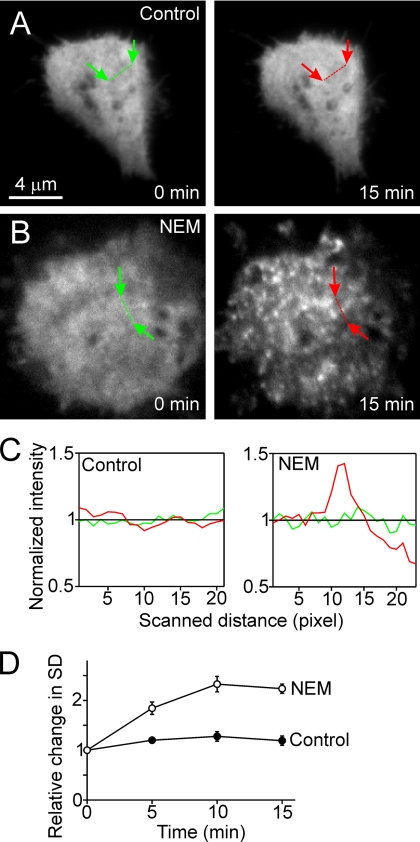
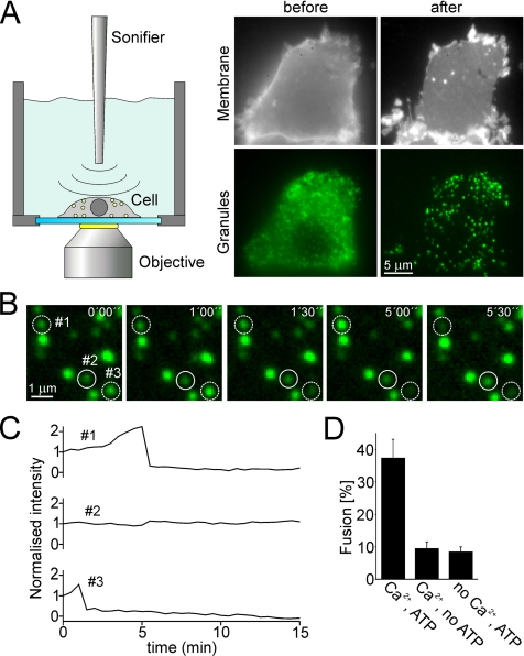
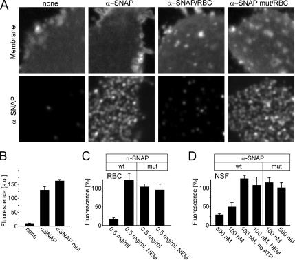
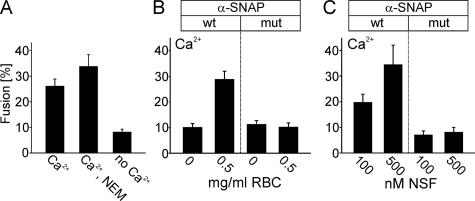
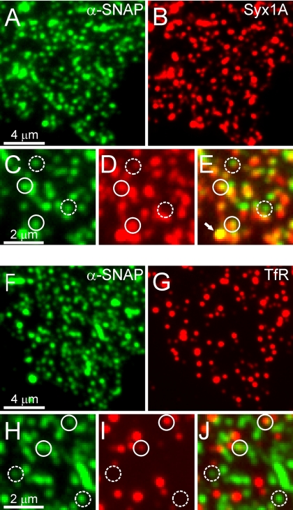
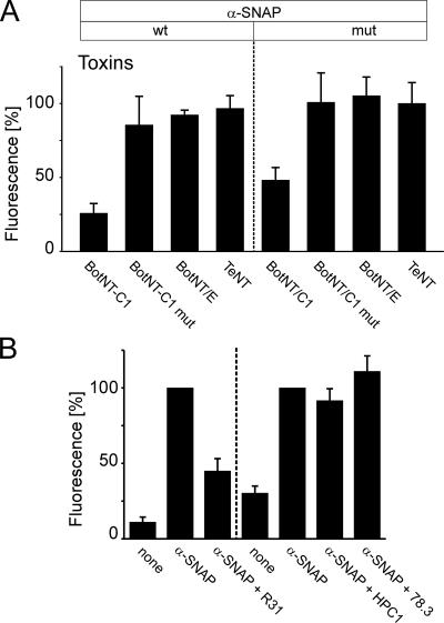
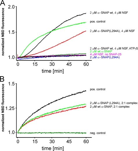
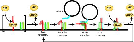
Regulated exocytosis in neurons and neuroendocrine cells requires the formation of a stable soluble N-ethylmaleimide-sensitive factor attachment protein receptor (SNARE) complex consisting of synaptobrevin-2/vesicle-associated membrane protein 2, synaptosome-associated protein of 25 kDa (SNAP-25), and syntaxin 1. This complex is subsequently disassembled by the concerted action of alpha-SNAP and the ATPases associated with different cellular activities-ATPase N-ethylmaleimide-sensitive factor (NSF). We report that NSF inhibition causes accumulation of alpha-SNAP in clusters on plasma membranes. Clustering is mediated by the binding of alpha-SNAP to uncomplexed syntaxin, because cleavage of syntaxin with botulinum neurotoxin C1 or competition by using antibodies against syntaxin SNARE motif abolishes clustering. Binding of alpha-SNAP potently inhibits Ca(2+)-dependent exocytosis of secretory granules and SNARE-mediated liposome fusion. Membrane clustering and inhibition of both exocytosis and liposome fusion are counteracted by NSF but not when an alpha-SNAP mutant defective in NSF activation is used. We conclude that alpha-SNAP inhibits exocytosis by binding to the syntaxin SNARE motif and in turn prevents SNARE assembly, revealing an unexpected site of action for alpha-SNAP in the SNARE cycle that drives exocytotic membrane fusion.


Figures

Figure 1.
Figure 1.
Recruitment of α-SNAP to the plasma membrane after NSF inactivation in vivo. (A and B) TIRF images of PC-12 cells expressing GFP-labeled α-SNAP. Cells were imaged for 15 min, taking one image per minute. Images for t = 0 and t = 15 min are shown. (A) Control, homogenous distribution of subplasmalemmal α-SNAP remains unchanged. (B) Treatment with 1 mM NEM causes α-SNAP to concentrate in plasmalemmal spots. Using linescans, the standard deviations of pixel intensities were determined (see Materials and Methods for details; see C for linescans normalized to average intensity) at t = 0 (green linescans) and t = 5, 10, and 15 min (red linescans). (D) Morphological alterations were expressed as relative changes in the SD of pixel intensities.
Figure 2.
Figure 2.
Ca2+-dependent exocytosis of secretory granules by using a membrane sheet-based cell-free assay. (A) Membrane sheets generated by sonication of PC12 cells retain docked secretory granules. Cells expressing the secretory granule marker NPY-GFP were grown on glass-coverslips and mounted on the microscope stage. GFP-labeled cells were selected and ruptured by brief pulses of ultrasound, resulting in a flat plasma membrane sheets with numerous green dots. Top, staining of the plasma membrane of a PC-12 cell before and after rupture with the lipophilic dye TMA-DPH. Bottom, GFP-channel showing secretory granules. (B) Granules docked to a membrane sheet undergo Ca2+-dependent exocytosis. Membrane sheet was preincubated for 5 min in an ATP-containing and calcium-free solution followed by the addition of ∼35 μM free calcium to trigger exocytosis (start at t = 0). Images were acquired every 30 s for 15 min. Exemplary images (time as indicated) show that the fluorescence intensity either changed (dashed circles) or remained constant (continuous circle). (C) Intensity traces of the granules encircled in B. Granules were scored as having undergone exocytosis when the drop of fluorescence intensity between two consecutive images exceeded 25%. (D) Exocytosis is dependent on the presence of Ca2+ and ATP in the triggering phase. Exocytotic membrane fusion was calculated by relating the number of granules scored positive for exocytosis during the 15 min stimulation phase to the number of granules present in the first image. Values are given as mean ± SEM (n = 9–20 membrane sheets for each condition recorded in at least three independent experiments). SEM denotes the SE of measurement.
Figure 3.
Figure 3.
Membrane sheets contain NSF-sensitive binding sites for α-SNAP. (A) Membrane sheets incubated for 5 min with recombinant α-SNAP or α-SNAPL294A were briefly washed, fixed, and immunostained for α-SNAP (bottom). Top, membrane sheets visualized using TMA-DPH (also see Figure 1A). Where indicated, RBC was included in the incubation. (B) Quantification of immunofluorescence intensity in the absence or presence of recombinant α-SNAP or α-SNAPL294A. For better comparison, in the following experiments (C and D; also see Figure 5) values were normalized to the corresponding immunoreactivity stainings in B. (C and D) Binding of α-SNAP (wt) but not of α-SNAPL294A (mut) is prevented by the inclusion of either RBC (C) or purified NSF (D). Addition of NEM (1 mM) or omission of ATP, both known to inactivate NSF, blocked the interference with α-SNAP binding by both rat brain cytosol and purified NSF. The concentrations of RBC (milligrams of protein per milliliter) and recombinant NSF (nanomolar) is given at the bottom of the columns. Values are given as mean ± SEM (n = 3–14 independent experiments, with 35–169 individual membrane sheets analyzed for each experiment).
Figure 4.
Figure 4.
Exocytosis is independent of NSF, but it is inhibited by α-SNAP. All experiments were performed as in Figure 2D, with ATP present in the preincubation and triggering phase and Ca2+ added during the triggering phase. All other additions were present only in the preincubation phase. (A) Preincubation of membrane sheets with 1 mM NEM does not inhibit exocytosis. (B and C) Preincubation with 2 μM α-SNAP (wt) or α-SNAPL294A (mut) inhibits Ca2+-dependent exocytosis. Inhibition by wild-type but not mutant α-SNAP is prevented by the inclusion of either RBC (B) or purified NSF (C). Values are given as mean ± SEM (n = 9–18 membrane sheets for each condition).
Figure 5.
Figure 5.
Syntaxin 1 clusters colocalize with α-SNAP binding sites. Membrane sheets were incubated with wild-type α-SNAP as given in Figure 3, and then they were fixed and double labeled for α-SNAP and syntaxin 1 (A–E) or transferrin receptor (TfR) (F–J). (C–E and H–J) Magnified views from A and B and F and G, respectively. Circles were placed on immunoreactive spot in the α-SNAP channel and transferred to identical pixel locations in the corresponding red channels. Immunoreactive spots were rated as colocalized according to Lang et al. (2002). After correction for accidental colocalization, we obtained 58 ± 2% specifically colocalizing spots for α-SNAP/syntaxin 1 and 9 ± 2% for α-SNAP/TfR (n = 10 membrane sheets analyzed per condition, with 50–200 spots analyzed on each membrane sheet; all values mean ± SEM). Solid circles indicate colocalizing, dashed circles noncolocalizing spots. The arrow in E indicates a fluorescent bead visible in all fluorescence channels that was added to the sample and used as a spatial reference for image alignment.
Figure 6.
Figure 6.
A-SNAP binds to the SNARE-motif of synatxin 1. (A) A-SNAP binding requires syntaxin 1 but not SNAP-25 or synaptobrevin. Membrane sheets were incubated for 5 min with 2 μM of either the wt- or mutant form of α-SNAP. Where indicated, solutions contained in addition 2 μM purified light chains of either BoNT/C1 cleaving syntaxin 1, BoNT/C1mut (inactive form of BoNT/C1 carrying the mutation E230A), BoNT/E cleaving SNAP-25, or TeNT cleaving synaptobrevin. After brief washing, membrane sheets were processed for immunostaining and analyzed as shown in Figure 3, C and D). Values are given as mean ± SEM (n = 3–4 independent experiments, with 70–120 individual [mean = 106] membrane sheets analyzed for each experiment). (B) Antibodies directed against the SNARE-motif of syntaxin inhibit binding of α-SNAP. Membrane sheets were incubated for 15 min with anti-syntaxin 1 antibodies, washed twice with PBS and followed by 5-min incubation with 2 μM recombinant wild-type α-SNAP. The sheets were then washed, fixed, and immunolabeled for α-SNAP. The antibodies used for preincubation were R31 (polyclonal rabbit antiserum recognizing both the N-terminal domain and the SNARE motif) and HPC1 and Cl 78.3 (independently raised monoclonal antibodies specific for the N-terminal Habc-domain). For the detection of α-SNAP, we used either a monoclonal (Cl 77.2, left) or a polyclonal rabbit antibody (R34, right). In all experiments, fluorescence values were normalized to the immunoreactivity of membrane-bound, recombinant α-SNAP without prior anti-syntaxin 1 antibody treatment. Values are given as mean ± SEM (n = 6–7 independent experiments, with a minimum of 10–144 individual membrane sheets analyzed for each experiment).
Figure 7.
Figure 7.
Inhibition of SNARE-mediated proteoliposome fusion by α-SNAP. Fusion was measured as an increase of NBD fluorescence by using a lipid dequenching assay. Donor liposomes were reconstituted with an N-terminally truncated version of syntaxin 1 (2 μM, residues 183–288) (A) or with a preformed complex of syntaxin 1 (same construct as before) and SNAP-25 (B). Acceptor liposomes contained 10 μM synaptobrevin 2. If syntaxin-containing liposomes were used as donor, the liposomes were combined and preincubated for 10 min at 30°C. Where indicated, solutions contained in addition α-SNAP, α-SNAPL294A, NSF, or ATPγS, and the reaction was started by addition of 10 μM soluble SNAP-25a. (t = 0, reference point for normalization of the signal). In B, donor liposomes contained a preformed syntaxin-SNAP-25 complex, and the reaction was started by mixing donor and acceptor liposomes.
Figure 8.
Figure 8.
Model illustrating the sites of action for α-SNAP and NSF in the SNARE conformational cycle during exocytosis. A-SNAP binds with high affinity to cis-SNARE complexes and with lower affinity to free syntaxin. Under normal circumstances, the equilibrium is shifted toward the dissociated part of the cycle, with the steady-state concentration of both cis-complexes and α-SNAP/syntaxin complexes being low. However, if the SNAP–NSF system is imbalanced due to overexpression of α-SNAP or down-regulation/inhibition of NSF, free syntaxin rapidly becomes rate limiting, resulting in an inhibition of exocytosis. Please note that free syntaxin is organized in clusters (not shown in the figure), and it is not known whether α-SNAP exhibits any preference for free versus clustered syntaxin. See text for details.

References

    1. Avery J., Ellis D. J., Lang T., Holroyd P., Riedel D., Henderson R. M., Edwardson J. M., Jahn R. A cell-free system for regulated exocytosis in PC12 cells. J. Cell Biol. 2000;148:317–324. - PMC - PubMed
    1. Babcock M., Macleod G. T., Leither J., Pallanck L. Genetic analysis of soluble N-ethylmaleimide-sensitive factor attachment protein function in Drosophila reveals positive and negative secretory roles. J. Neurosci. 2004;24:3964–3973. - PMC - PubMed
    1. Banerjee A., Barry V. A., DasGupta B. R., Martin T. F. N-Ethylmaleimide-sensitive factor acts at a prefusion ATP-dependent step in Ca2+-activated exocytosis. J. Biol. Chem. 1996;271:20223–20226. - PubMed
    1. Barnard R. J., Morgan A., Burgoyne R. D. Stimulation of NSF ATPase activity by alpha-SNAP is required for SNARE complex disassembly and exocytosis. J. Cell Biol. 1997;139:875–883. - PMC - PubMed
    1. Barnstable C. J., Hofstein R., Akagawa K. A marker of early amacrine cell development in rat retina. Brain Res. 1985;352:286–290. - PubMed

MeSH terms

Substances

LinkOut - more resources