. 2008 Mar;19(3):785-96.
doi: 10.1091/mbc.e07-05-0497. Epub 2007 Dec 19.

DYC-1, a protein functionally linked to dystrophin in Caenorhabditis elegans is associated with the dense body, where it interacts with the muscle LIM domain protein ZYX-1

Affiliations

DYC-1, a protein functionally linked to dystrophin in Caenorhabditis elegans is associated with the dense body, where it interacts with the muscle LIM domain protein ZYX-1

Claire Lecroisey et al. Mol Biol Cell. 2008 Mar.

Abstract

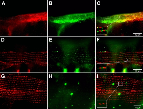
In Caenorhabditis elegans, mutations of the dystrophin homologue, dys-1, produce a peculiar behavioral phenotype (hyperactivity and a tendency to hypercontract). In a sensitized genetic background, dys-1 mutations also lead to muscle necrosis. The dyc-1 gene was previously identified in a genetic screen because its mutation leads to the same phenotype as dys-1, suggesting that the two genes are functionally linked. Here, we report the detailed characterization of the dyc-1 gene. dyc-1 encodes two isoforms, which are expressed in neurons and muscles. Isoform-specific RNAi experiments show that the absence of the muscle isoform, and not that of the neuronal isoform, is responsible for the dyc-1 mutant phenotype. In the sarcomere, the DYC-1 protein is localized at the edges of the dense body, the nematode muscle adhesion structure where actin filaments are anchored and linked to the sarcolemma. In yeast two-hybrid assays, DYC-1 interacts with ZYX-1, the homologue of the vertebrate focal adhesion LIM domain protein zyxin. ZYX-1 localizes at dense bodies and M-lines as well as in the nucleus of C. elegans striated muscles. The DYC-1 protein possesses a highly conserved 19 amino acid sequence, which is involved in the interaction with ZYX-1 and which is sufficient for addressing DYC-1 to the dense body. Altogether our findings indicate that DYC-1 may be involved in dense body function and stability. This, taken together with the functional link between the C. elegans DYC-1 and DYS-1 proteins, furthermore suggests a requirement of dystrophin function at this structure. As the dense body shares functional similarity with both the vertebrate Z-disk and the costamere, we therefore postulate that disruption of muscle cell adhesion structures might be the primary event of muscle degeneration occurring in the absence of dystrophin, in C. elegans as well as vertebrates.


Figures

Figure 1.
Figure 1.
Structure of the dyc-1 gene and constructs used in this study. Top, cosmids C33G3 and C04C11 that overlap with the dyc-1 gene. Middle, genomic organization of the dyc-1 gene. Rectangles indicate the different exons. The dyc-1 gene encodes two different isoforms: a short protein of 793 amino acids called DYC-1S and a long 887 amino acid protein called DYC-1L. These isoforms are generated from alternative promoters, DYC-1S is expressed in muscles and DYC-1L in neurons. Both DYC-1 proteins contain two regions of strong homology to the human protein CAPON (indicated in blue). Isofom-specific regions used in the RNAi experiments are shown as red bars. The respective positions of the dyc-1(cx5) and the dyc-1(cx32) mutations are indicated (arrows). Bottom, structure of two transgenes cited in this article. Note that the dyc-1:gfpVI transgene does not contain the neuronal promoter, whereas dyc-1:gfpX contains both the neuronal and the muscular promoter.
Figure 2.
Figure 2.
Muscular and neuronal isoform-specific RNAi experiments. (A) Control animal expressing the dyc-1:gfpX transgene fed with bacteria containing the empty plasmid (L4440). Arrow shows the muscular expression of the transgene and arrowhead indicates the cell body of a neuron expressing the transgene. (B) Transgenic animals fed with muscular isoform specific RNAi. Note the strong reduction of muscular GFP signal, whereas the neuronal signal remains intense (arrowhead). (C) Transgenic animals fed with neuronal isoform-specific RNAi. The arrowhead points to a neuron. Note that the neuronal GFP signal is slightly reduced but not completely abolished. The muscular GFP signal (arrow) seems not to differ from control animals.
Figure 3.
Figure 3.
Neuronal dyc-1 expression seen by reporter gene. Immunofluorescence images of wild-type animals carrying a dyc-1:gfpX transgene. Expression is detected in ∼10 neurons. A neuron cell body (SDQR) is visible in A (arrowhead). In A and B arrows indicate the punctate pattern along the axons.
Figure 4.
Figure 4.
Subcellular localization of DYC-1 protein in muscle cells. Immunofluorescence images of wild-type animals (A–F) and dyc-1(cx32) mutants (G–I). (A, D, and G) Dense body localization of the DEB-1/vinculin protein labeled with MH24 antibody (red). (B) Localization of the dyc-1:gfpX reporter gene (green, the signal is amplified with anti-GFP antibodies). (C) Merge of A and B. In the magnification of two dense bodies, the green GFP signal is seen at the edges of the red MH24 signal. (E) Localization of the DYC-1 protein labeled with purified anti-DYC-1 antibodies (green). (F) Merge of D and E with a magnification of two dense bodies. Note that as in C, the green (DYC-1) signal is located at the edges of dense bodies (red MH24 signal). (H) dyc-1(cx32) mutants stained with anti-DYC-1 antibodies. No specific signal can be detected. (I) Merge of G and H with a detail of two dense bodies showing the absence of DYC-1 staining.
Figure 5.
Figure 5.
Sarcomeric localization of the DYC-1 protein in immunoelectron microscopy. (A and B) Examples of cross-sections of dyc-1:gfpX expressing transgenic worms labeled with an anti-GFP antibody coupled to gold beads. Note that gold beads are preferentially located at the bottom of the dense body (arrows). Clusters of gold beads in the cytoplasm (arrowheads in A), are due to overexpression of the transgene. (C) Diagram summarizing quantification of gold beads according to their sarcomeric position. These results are the mean of two different counts. 72.34% of gold beads are located in the dense body region and 27.66% outside of it. Among the gold beads which are located in the dense body zone, 4.30% are located in the dense body (3.52% at the bottom and 0.78% on the middle) and 68.04% are located at the edges (between two dense bodies), with 43.70% of them at the bottom zone, 12.02% in the middle zone, and 12.32% in the top zone. (D) Detail of a dense body showing the limits of different zones considered for scoring gold bead locations. In this picture the section does not cross the dense body (otherwise it would appear in black). Gold beads located at this level are therefore classified as being at the edge of the dense body.
Figure 6.
Figure 6.
Subcellular localization of the ZYX-1 protein by reporter gene analysis. (A–H) Transgenic animals expressing a zyx-1:gfp construct. Body-wall muscles of animals labeled with anti-DEB-1 MH24 antibody (red in A and C) or anti-DYC-1 antibodies (red in D and F). (B and E) The muscle localization of the ZYX-1-GFP protein (green, the GFP signal was amplified with an anti-GFP antibody). ZYX-1-GFP localizes on dense bodies (arrowhead in B) and M-lines (arrow in B). (C and F) Merged images of A and B and of D and E, respectively, with details of two dense bodies. Note in C that the DEB-1 signal colocalizes with the ZYX-1-GFP signal on the dense body and in F that the DYC-1 signal is located at the edges of the ZYX-1-GFP labeling. (G) Localization of ZYX-1-GFP in the nucleus (arrowhead) of a body wall muscle cell. (H) Localization of ZYX-1-GFP in neurons (arrowheads).
Figure 7.
Figure 7.
DYC-1S interacts with ZYX-1 through its highly conserved 19 aa region. (A) Summary of results obtained with yeast two-hybrid assay. The different parts of DYC-1 and ZYX-1 proteins used in these assays are schematically indicated (top left). The table indicates the different plasmids used in these assays and the corresponding DYC-1 bait and ZYX-1 prey proteins. Pictures show plates inoculated with diploid yeasts expressing the different DYC-1S bait and ZYX-1 prey proteins. Medium without Leu and Trp serves as a positive control showing the presence of bait and prey plasmids in the tested diploid yeasts. On medium without His, Leu, and Trp, growth can occur only if the bait and the prey proteins interact. Note that negative control yeasts carrying either empty pACT2 or pAS2-1 plasmids do not grow on medium without His, Leu, and Trp. Interactions occur between the bait proteins DYC-1S (aa 9-793) and DYC-1S (aa 9-258) and the prey proteins ZYX-1 (aa 68-603) and ZYX-1 (aa 384-603; growth on medium without His, Leu, and Trp). Note that interaction is detected neither for the bait protein DYC-1S (aa 9-258 Δaa 54-81) carrying a deletion of aa 54-81, nor for the prey protein ZYX-1 (aa 384-528) carrying only the two first LIM domains of the ZYX-1 protein (no growth on medium without His, Leu, and Trp). (B) Western blotting of GST pulldown assays. HA-tagged ZYX-1 protein (aa 15-603) was detected using an anti-HA antibody after GST pulldown performed with the GST-tagged N-terminal end of DYC-1 protein (aa 9-258) but not with GST alone. The right lane shows ZYX-1-HA expression in the total COS-7 cell extract. A volume of 20 μl was analyzed on each lane. (C) Alignment of a DYC-1S conserved sequence. Ce, Caenorhabditis elegans,; Dm, Drosophila; Ag, Anopheles; Rn, Rat; Hs, humans. Note that the region of 19 aa (aa 58-76 of the DYC-1S protein), which is strictly conserved from Drosophila to humans (two conservative differences exist in C. elegans, red arrows).
Figure 8.
Figure 8.
Subcellular localization of GFP fused to the highly conserved stretch of the DYC-1S protein. (A) Dense body localization of DEB-1 protein labeled with MH24 antibody (red). (B) Localization of GFP (green) fused to DYC-1S (aa 54-81) containing the highly conserved stretch of 19 aa. (C) Merged image of A and B with the detail of two dense bodies. Note that the short sequence of the DYC-1S protein is sufficient to address GFP to the sarcomeric localization.
Figure 9.
Figure 9.
Working model of dystrophin-DYC-1-dense body interactions. Dystrophin (in green) is known to interact with the Dystroglycan (DG)–Sarcoglycan (SG) complex (blue) by its C-terminal end. DYS-1/dystrophin interacts with DYB-1/dystrobrevin, which in turn binds STN-1/syntrophin. PAT-2/α-integrin and PAT-3/β-integrin (yellow) anchors the dense body to the sarcolemma and the extracellular matrix. DYC-1 (purple) is located at the edges of the dense body and interacts via its conserved 19-aa stretch (small purple bead) with the third LIM domain of ZYX-1/zyxin (the three LIM domains of the Zyx-1 protein are represented as small red beads). ZYX-1 is also located in the nucleus of muscle cells where it might mediate signaling pathways. We posit that there is a direct or an indirect interaction between dystrophin and DYC-1, linking DYS-1/dystrophin to the dense body.

Similar articles

Cited by

References

    1. Ahn A. H., Kunkel L. M. The structural and functional diversity of dystrophin. Nat. Genet. 1993;3:283–291. - PubMed
    1. Barstead R. J., Waterston R. H. Vinculin is essential for muscle function in the nematode. J. Cell Biol. 1991;114:715–724. - PMC - PubMed
    1. Benian G. M., L'Hernault S. W., Morris M. E. Additional sequence complexity in the muscle gene, unc-22, and its encoded protein, twitchin, of Caenorhabditis elegans. Genetics. 1993;134:1097–1104. - PMC - PubMed
    1. Benian G. M., Tinley T. L., Tang X., Borodovsky M. The Caenorhabditis elegans gene unc-89, required fpr muscle M-line assembly, encodes a giant modular protein composed of Ig and signal transduction domains. J. Cell Biol. 1996;132:835–848. - PMC - PubMed
    1. Bessou C., Giugia J. B., Franks C. J., Holden-Dye L., Segalat L. Mutations in the Caenorhabditis elegans dystrophin-like gene dys-1 lead to hyperactivity and suggest a link with cholinergic transmission. Neurogenetics. 1998;2:61–72. - PubMed

Publication types

MeSH terms

LinkOut - more resources