Review
. 2008 Jan 1;5(1):4-10.
doi: 10.1513/pats.200704-049VS.

Embryological origin of airway smooth muscle

Affiliations
Review

Embryological origin of airway smooth muscle

Kameswara Rao Badri et al. Proc Am Thorac Soc. .

Abstract

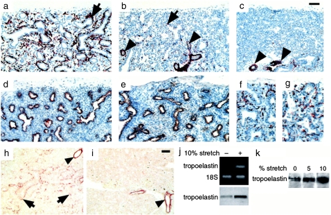
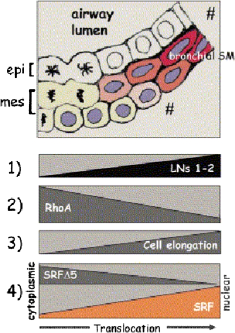
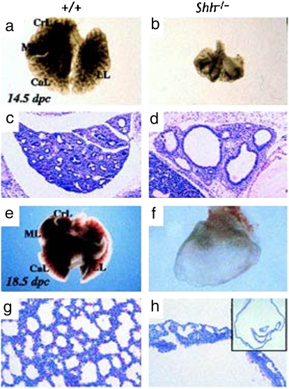
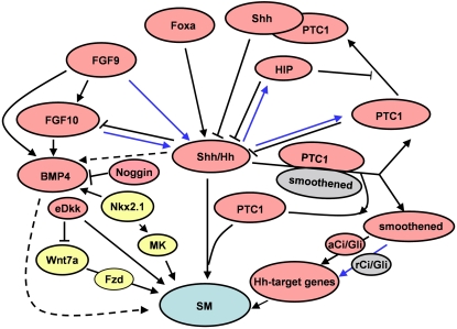
Airway smooth muscle (SM) develops from local mesenchymal cells located around the tips of growing epithelial buds. These cells gradually displace from distal to proximal position alongside the bronchial tree, elongate, and begin to synthesize SM-specific proteins. Mechanical tension (either generated by cell spreading/elongation or stretch), as well as epithelial paracrine factors, regulates the process of bronchial myogenesis. The specific roles of many of these paracrine factors during normal lung development are currently unknown. It is also unknown how and if mechanical and paracrine signals integrate into a common myogenic pathway. Furthermore, as with vascular SM and other types of visceral SM, we are just beginning to elucidate the intracellular signaling pathways and the genetic program that controls lung myogenesis. Here we present what we have learned so far about the embryogenesis of bronchial muscle.


Figures

<b>Figure 1.</b>
Figure 1.
Embryonic whole lung explants cultured in the presence of extra- and intra-luminal dextran (volume expander) and immunostained for smooth muscle (SM) α-actin. (a) SM α-actin in control explants cultured without dextran (unmodified pressure). Some noticeable bronchial SM development is present (arrows). (b) SM α-actin in lung explants cultured with 5% dextran inside airways (increased pressure) shows hyperplastic bronchial muscle (arrow). (c) No bronchial myogenesis in lung explants cultured with 1% dextran in the culture medium (eliminated pressure). Bar = 100 μm. Reprinted by permission from Reference .
<b>Figure 2.</b>
Figure 2.
Immunohistochemistry showing paucity of bronchial SM cells in human fetal hypoplastic lungs. Shown are histologic sections from (a) normal lung, (b) hypoplastic lung caused by oligohydramnion, and (c) hypoplastic lung caused by diaphragmatic hernia, all at 22 weeks of gestation, immunostained for SM α-actin. There is significant decrease in bronchial SM cells (arrows) in the hypoplastic lungs (b and c), particularly in those compressed by intrathoracic herniation of abdominal viscerae due to diaphragmatic hernia (c). The vascular musculature is unaffected (arrowheads). In the same hypoplastic lung shown in b, the epithelial cells, immunostained for cytokeratins (e), and the endothelial cells, immunostained for PECAM-1 (g), show no changes compared with controls (d and f). h and i demonstrate immunohistochemistry showing decrease in tropoelastin deposition in human hypoplastic lungs. (h) Histologic sections from normal lung at 20 weeks of gestation demonstrate tropoelastin deposition around bronchi and bronchioli (arrows). (i) Histologic sections from same age hypoplastic lung reveal essentially no tropoelastin deposition, with the exception of vascular SM that shows no changes in tropoelastin when compared with controls (arrowheads). Bar = 60 μm in ae, h, and i, and 100 μm in f and g. RT-PCR and immunoblot show stretch-induced upregulation of tropoelastin expression in mouse lung embryonic mesenchymal cells undergoing myogenic differentiation. (k) Immunoblot shows stretch-induced upregulation of tropoelastin synthesis in human lung embryonic mesenchymal cells undergoing myogenic differentiation. Reprinted by permission from Reference .
<b>Figure 3.</b>
Figure 3.
New epithelial–mesenchymal contacts at the tips of growing airway buds induce laminin (LN)-1 expression and deposition at the epithelial–mesenchymal interface (1). LN-1 is a powerful inhibitor of RhoA activity (2). Because high RhoA activity maintains the undifferentiated mesenchymal cells' round shape, its decrease allows for initial cell elongation (3). Cell elongation then switches the synthesis of LN-2 (1), which is an even more powerful inhibitor of RhoA. A positive feedback loop is created between LN-2, RhoA inhibition, and cell elongation (1–3). In the round, undifferentiated mesenchymal cells serum response factor (SRF) and SRFΔ5 are present in the nucleus and cytoplasm (4). Upon cell elongation, the cytoplasmic SRF isoform translocates gradually to the nucleus, whereas SRFΔ5 disappears (4). The increment of SRF plus the disappearance of SRFΔ5 contribute to the initiation of bronchial myogenesis. Reprinted by permission from Reference .
<b>Figure 4.</b>
Figure 4.
Shh is essential in lung development. Whole mount view of (a, e) wild-type and (b, f) Shh−/− mutant lungs at (a, b) 14.5 and (e, f) 18.5 days after conception. Wild type shows characteristic lobulation (CrL, cranial lobe; ML, middle lobe; CaL, caudal lobe; LL, left lobe), which is not present in the mutant. Note that the mutant lung at 18.5 days after conception is reduced to a dilated sac (h, inset). (c, d, g, and h) Hematoxylin and eosin–stained sections of (c, g) wild type and (d, h) mutant lungs at (c, d) 14.5 and (g, h) 18.5 days after conception. Reprinted by permission from Reference .
<b>Figure 5.</b>
Figure 5.
A schematic representation of signaling pathways in lung smooth muscle myogenesis. Blue lines indicate a negative regulation on Shh-mediated smooth muscle myogenesis. Dotted lines represent relationships that are not yet clear. Yellow represents involvement in vascular smooth muscle myogenesis only. Gray represents repressed protein or complex. Shh, sonic hedgehog; Hh, hedgehog; PTC1, patched-1; HIP, hedgehog-interacting protein; aCi/Gli, Ci/Gli activator; rCi/Gli, Ci/Gli repressor; Foxa, Forkhead box-a; FGF, fibroblast growth factor; BMP4, bone morphogenetic protein-4; eDkk, epithelial Dickkopf-1; Fzd, Frizzled; MK, midkine, SM, smooth muscle myogenesis.

References

    1. Sparrow MP, Lamb JP. Ontogeny of airway smooth muscle: structure, innervation, myogenesis and function in the fetal lung. Respir Physiolo Neurobiol 2003;137:361–372. - PubMed
    1. Roman J, McDonald JA. Expression of fibronectin, the integrin alpha 5, and alpha-smooth muscle actin in heart and lung development. Am J Respir Cell Mol Biol 1992;6:472–480. - PubMed
    1. McHugh KM. Molecular analysis of smooth muscle development in the mouse. Dev Dyn 1995;204:278–290. - PubMed
    1. Li L, Miano JM, Cserjesi P, Olson EN. Sm22 alpha, a marker of adult smooth muscle, is expressed in multiple myogenic lineages during embryogenesis. Circ Res 1996;78:188–195. - PubMed
    1. Mitchell HW, Sparrow MP, Tagliaferri RP. Inhibitory and excitatory responses to field stimulation in fetal and adult pig airway. Pediatr Res 1990;28:69–74. - PubMed

Publication types

LinkOut - more resources