. 2008 Feb;14(2):367-80.
doi: 10.1261/rna.696508. Epub 2007 Dec 19.

Factor requirements for translation initiation on the Simian picornavirus internal ribosomal entry site

Affiliations

Factor requirements for translation initiation on the Simian picornavirus internal ribosomal entry site

Sylvain de Breyne et al. RNA. 2008 Feb.

Abstract

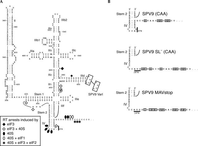
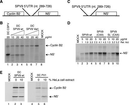
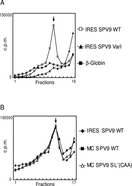
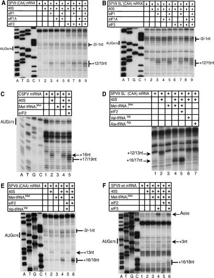
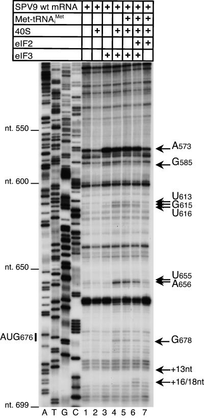
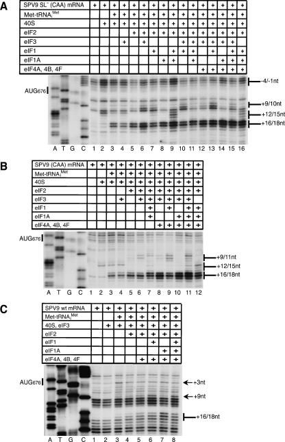
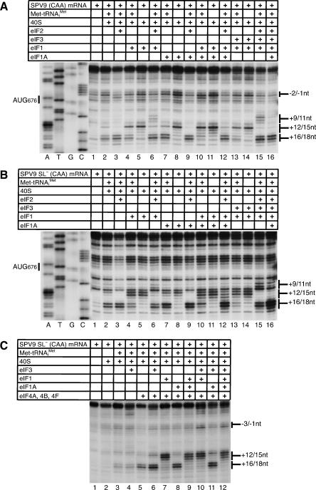
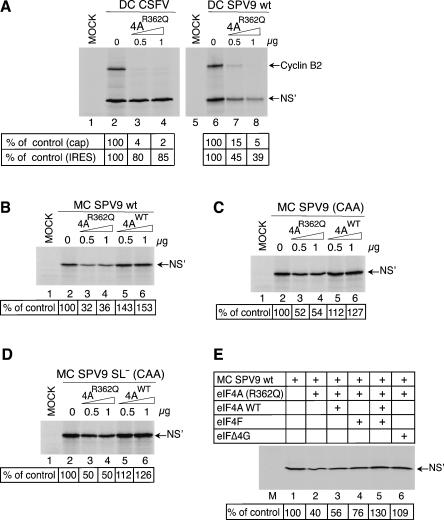
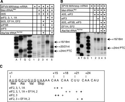
The Simian picornavirus type 9 (SPV9) 5'-untranslated region (5' UTR) has been predicted to contain an internal ribosomal entry site (IRES) with structural elements that resemble domains of hepacivirus/pestivirus (HP) IRESs. In vitro reconstitution of initiation confirmed that this 5' UTR contains an IRES and revealed that it has both functional similarities and differences compared to HP IRESs. Like HP IRESs, the SPV9 IRES bound directly to 40S subunits and eukaryotic initiation factor (eIF) 3, depended on the conserved domain IIId for ribosomal binding and consequently for function, and additionally required eIF2/initiator tRNA to yield 48S complexes that formed elongation-competent 80S ribosomes in the presence of eIF5, eIF5B, and 60S subunits. Toeprinting analysis revealed that eIF1A stabilized 48S complexes, whereas eIF1 induced conformational changes in the 40S subunit, likely corresponding to partial opening of the entry latch of the mRNA-binding channel, that were exacerbated by eIF3 and suppressed by eIF1A. The SPV9 IRES differed from HP IRESs in that its function was enhanced by eIF4A/eIF4F when the IRES was adjacent to the wild-type coding sequence, but was less affected by these factors or by a dominant negative eIF4A mutant when potentially less structured coding sequences were present. Exceptionally, this IRES promoted binding of initiator tRNA to the initiation codon in the P site of 40S subunits independently of eIF2. Although these 40S/IRES/tRNA complexes could not form active 80S ribosomes, this constitutes a second difference between the SPV9 and HP IRESs. eIF1 destabilized the eIF2-independent ribosomal binding of initiator tRNA.


Figures

FIGURE 1.
FIGURE 1.
(A) Model of the secondary and tertiary structure of the Simian picornavirus type 9 IRES (nucleotides 308–697) (Hellen and de Breyne 2007). The nomenclature of domains is based on proposals for the HCV IRES (Honda et al. 1996). The initiation codon AUG676 and the substitutions GGGG593–596AAAA in SPV9 (Var1) are indicated. Sites at which primer extension was arrested or enhanced by binding of eIF3, of 40S subunits, of 40S subunits with eIF3 or eIF1, and of the 48S complex to the IRES are indicated by symbols (shown at the lower left). (B) Sequences flanking the initiation codon of SPV9 (CAA), SPV9 SL (CAA), and SPV9 MAVstop variants, in which substitutions relative to the wild-type coding sequence are boxed.
FIGURE 2.
FIGURE 2.
Effect of cis-acting elements on SPV9 IRES function. (A) Schematic representation of dicistronic mRNAs containing cyclin B2 and influenza NS′ cistrons separated by either CSFV nucleotides 1–442, SPV9 (wt), or SPV9 (Var1). (C) Schematic representation of monocistronic mRNAs containing the NS′ cistron preceded by SPV9 (wt), SPV9 (CAA), or SPV9 SL (CAA). (B,D) Translation of dicistronic (B) and monocistronic mRNAs (D) at the indicated concentrations in RRL containing [35S]methionine. Products were analyzed by gel electrophoresis and autoradiography. SPV9 IRES-mediated initiation (D) was quantified using a Molecular Dynamics PhosphoImager relative to translation of SPV9 (wt) mRNA (5 μg/mL) (lane 2), which was defined as 1. (E) The influence of a cytoplasmic S10 extract added to RRL at the indicated proportions on translation of DC SPV9 (wt) mRNA (lanes 2,3) and DC PV1 mRNA (lanes 5,6) was quantified using a Molecular Dynamics PhosphorImager after separation of translation products by gel electrophoresis. Lanes labeled “MOCK” correspond to RRL incubated without exogenous mRNA.
FIGURE 3.
FIGURE 3.
Factor-independent binding of 40S subunits to the SPV9 IRES. Ribosomal complexes were assembled by incubating (A) [32P]UTP-labeled SPV9 (wt), SPV9 (Var1), or β-globin mRNAs or (B) [32P]UTP-labeled SPV9 (wt), MC SPV9 (wt), or MC SPV9 SL (CAA) mRNAs (as indicated) with 40S subunits. Ribosomal complexes were analyzed by centrifugation through a 10%–30% sucrose density gradient. Sedimentation was from right to left. The position of binary IRES–40S subunit complexes is indicated. Fractions from upper parts of the gradient have been omitted for greater clarity.
FIGURE 4.
FIGURE 4.
eIF2-independent formation of 48S initiation complexes. MC SPV9 (CAA) (A,E), MC SPV9 SL (CAA) (B,D), DC CSFV (C), and SPV9 (wt) mRNA (F) were incubated with 40S subunits, eIF2, eIF3, Met-tRNAi Met, Ala-tRNAAla, and Val-tRNAVal as indicated above each panel. Reaction conditions are described in Materials and Methods. Extension by AMV-RT of a primer annealed to the NS′ cistron was arrested at sites indicated on the right (A,B,D–F) or left (D) as positions relative to the A (position +1) of the SPV9 initiation codon (A,B,D,E) or the CSFV initiation codon. Reference lanes A, T, G, and C depict (A,B,E,F) SPV9 (CAA) and (C) CSFV sequences. For clarity, only a portion of each gel is shown.
FIGURE 5.
FIGURE 5.
Influence of eIF3 on the IRES–40S subunit complex. DC SPV9 (wt) mRNA was incubated with 40S subunits, eIF2, eIF3, and Met-tRNAi Met as indicated, in standard conditions. Extension by AMV-RT of a primer annealed to the NS′ cistron was arrested at sites indicated on the right either as nucleotides in the SPV9 5′ UTR or as positions relative to the A (position +1) of the initiation codon. Reference lanes A, T, G, and C depict the SPV9 (CAA) sequence.
FIGURE 6.
FIGURE 6.
The influence of initiation factors and proximal coding sequences on 48S complex formation on the SPV9 IRES. (A) MC SPV9 SL (CAA), (B) MC SPV9 (CAA), and (C) DC SPV9 (wt) mRNA were incubated with 40S subunits, eIF2, eIF3, eIF1, eIF1A, eIF4A, eIF4B, eIF4F, and Met-tRNAi Met as indicated, in standard conditions. Extension by AMV-RT of a primer annealed to the NS′ cistron was arrested at sites indicated on the right as positions relative to the A (position +1) of the initiation codon. Reference lanes A, T, G, and C depict the SPV9 (CAA) sequence.
FIGURE 7.
FIGURE 7.
Effect of eIF1 and eIF1A on 48S complex formation. MC SPV9 (CAA) (A) and MC SPV9 SL (CAA) (B,C) mRNAs were incubated with 40S subunits, eIF2, eIF3, eIF1, eIF1A, eIF4A, eIF4B, eIF4F, and Met-tRNAi Met as indicated, in standard conditions. Extension by AMV-RT of a primer annealed to the NS′ cistron was arrested at sites indicated on the right as positions relative to the A (position +1) of the initiation codon. Reference lanes A, T, G, and C depict the SPV9 (CAA) sequence.
FIGURE 8.
FIGURE 8.
The influence of proximal coding sequences on the sensitivity of SPV9 IRES-mediated translation to inhibition by trans-dominant eIF4AR362Q. (A) RRL was preincubated alone (lanes 2,6) or with the indicated amounts (μg) of eIF4AR362Q mutant (lanes 3,4,7,8) for 5 min at 30°C and then incubated for 55 min at 30°C without exogenous mRNA (lanes 1,5), with DC CSFV mRNA (lanes 2–4) or DC SPV9 (wt) mRNA (lanes 6–8). (B–D) RRL was pre-incubated alone (lanes 1,2) or with the indicated amounts (μg) of eIF4Awt (lanes 3,4) or eIF4AR362Q mutant (lanes 5,6) for 5 min at 30°C and then incubated for 55 min at 30°C with (A) DC CSFV, (B) DC SPV9 (wt), (B) MC SPV9 (wt), (C) MC SPV9 (CAA), (D) MC SPV9 SL (CAA) mRNAs (10 μg/mL), or (B–D, lane 1) without exogenous mRNA. (E) MC SPV9 (wt) mRNA (10 μg/mL) was incubated in pretreated RRL without (lane 1) or with 1 μg eIF4AR362Q mutant (lanes 2–6) and with 1 μg of eIF4Awt, eIF4F, or eIFΔ4G as indicated for 55 min at 30°C. (Lane M) Incubation of RRL without exogenous mRNA or factors for 55 min at 30°C. Translation products were visualized by autoradiography after electrophoresis on NuPAGE 4%–12% Bis-Tris-Gel. SPV9 IRES-mediated initiation was quantified using a Molecular Dynamics PhosphorImager relative to translation in the absence of exogenous eIF4A, which was defined as 100%.
FIGURE 9.
FIGURE 9.
Factor requirements for assembly of 48S complexes that can form elongation-competent ribosomes. (A,B) MC SPV9 MAVstop mRNA was incubated for 10 min at 37°C with 40S subunits, eIFs 1, 1A, 2 and 3, and Met-tRNAi Met as indicated to assemble 48S complexes. 80S ribosomes were formed by incubating these 48S complexes with eIF5, eIF5B, and 60S subunits for 10 min at 37°C, and were programmed to complete two cycles of codon-dependent elongation by incubation with eEF1H, eEF2, Ala-tRNAAla, and Val-tRNAVal for 10 min at 37°C. A primer annealed to the NS′ cistron and extended with AMV-RT was arrested at sites indicated on the right as positions relative to the A (position +1) of the AUG codon. Reference lanes A, T, G, and C depict (panel A) SPV9 (CAA) and (panel B) the SPV9 MAVstop sequences. PTC refers to the ribosomal pretermination complex, which contains peptidyl-tRNA in the P site and the stop codon in the A site. (C) Summary of the positions of ribosomal complexes assembled on SPV9 MAVstop mRNA in the presence of initiation and elongation factors as indicated. The positions of toeprints relative to nucleotide +1 of the AUG codon in the P site before translocation are indicated schematically below the SPV9 MAVstop sequence.

References

    1. Algire, M.A., Maag, D., Lorsch, J.R. Pi release from eIF2, not GTP hydrolysis, is the step controlled by start-site selection during eukaryotic translation initiation. Mol. Cell. 2005;20:251–262. - PubMed
    1. Alkalaeva, E.Z., Pisarev, A.V., Frolova, L.Y., Kisselev, L.L., Pestova, T.V. In vitro reconstitution of eukaryotic translation reveals cooperativity between release factors eRF1 and eRF3. Cell. 2006;125:1125–1136. - PubMed
    1. Battiste, J.L., Pestova, T.V., Hellen, C.U.T., Wagner, G. The eIF1A solution structure reveals a large RNA-binding surface important for scanning function. Mol. Cell. 2000;5:109–119. - PubMed
    1. Bordeleau, M.E., Mori, A., Oberer, M., Lindqvist, L., Chard, L.S., Higa, T., Belsham, G.J., Wagner, G., Tanaka, J., Pelletier, J. Functional characterization of IRESes by an inhibitor of the RNA helicase eIF4A. Nat. Chem. Biol. 2006;2:213–220. - PubMed
    1. Carter, A.P., Clemons W.M., Jr, Brodersen, D.E., Morgan-Warren, R.J., Hartsch, T., Wimberly, B.T., Ramakrishnan, V. Crystal structure of an initiation factor bound to the 30S ribosomal subunit. Science. 2001;291:498–501. - PubMed

Publication types

MeSH terms