. 2008 Mar;82(5):2385-93.
doi: 10.1128/JVI.01946-07. Epub 2007 Dec 19.

The DNase of gammaherpesviruses impairs recognition by virus-specific CD8+ T cells through an additional host shutoff function

Affiliations

The DNase of gammaherpesviruses impairs recognition by virus-specific CD8+ T cells through an additional host shutoff function

Jianmin Zuo et al. J Virol. 2008 Mar.

Abstract

The DNase/alkaline exonuclease (AE) genes are well conserved in all herpesvirus families, but recent studies have shown that the AE proteins of gammaherpesviruses such as Epstein-Barr virus (EBV) and Kaposi's sarcoma-associated herpesvirus (KSHV) exhibit an additional function which shuts down host protein synthesis. One correlate of this additional shutoff function is that levels of cell surface HLA molecules are downregulated, raising the possibility that shutoff/AE genes of gammaherpesviruses might contribute to viral immune evasion. In this study, we show that both BGLF5 (EBV) and SOX (KSHV) shutoff/AE proteins do indeed impair the ability of virus-specific CD8+ T-cell clones to recognize endogenous antigen via HLA class I. Random mutagenesis of the BGLF5 gene enabled us to genetically separate the shutoff and AE functions and to demonstrate that the shutoff function was the critical factor determining whether BGLF5 mutants can impair T-cell recognition. These data provide further evidence that EBV has multiple mechanisms to modulate HLA class I-restricted T-cell responses, thus enabling the virus to replicate and persist in the immune-competent host.


Figures
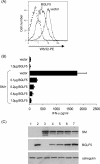
FIG. 1.
FIG. 1.
BGLF5 early antigen downregulates HLA class I expression in 293 cells. (A) The kinetics of BGLF5 expression following synchronous induction of the lytic cycle in EBV-positive Akata cells was analyzed by Western blotting with rabbit polyclonal antiserum to BGLF5 (middle blot). Expression of the immediate-early BZLF1 protein (top blot) was detected with the BZ.1 mouse monoclonal antibody, and the late p18 VCA protein (bottom blot) was detected with rabbit antiserum to BFRF3. (B) 293 cells transfected with pCDNA3-BGLF5-IRES-nlsGFP or the control pCDNA3-IRES-nlsGFP vector were analyzed at 48 h after transfection for expression of HLA class I expression at the cell surface. Cells were stained with PE-conjugated W6/32 antibody and analyzed by flow cytometry. (C) 293 cells transfected with pCDNA3-BGLF5-IRES-nlsGFP or the control pCDNA3-IRES-nlsGFP vector were subjected to fluorescence-activated cell sorting at 24 h after transfection to isolate GFP+ cells. Total lysates from 2 × 10⁵ sorted cells were analyzed by SDS-PAGE and Western blotting using a rabbit polyclonal antiserum specific for BGLF5 or the murine HC10 MAb specific to HLA class I heavy chains.
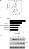
FIG. 2.
FIG. 2.
EBV BGLF5 can inhibit the T-cell recognition of 293 cells. (A) 293 cells were transfected with pCDNA3-BGLF5-IRES-nlsGFP or the control pCDNA3-IRES-nlsGFP vector. At 48 h after transfection, HLA class I expression at the surface of transfected cells was visualized by flow cytometry. The solid line histograms depict the surface HLA class I staining with PE-conjugated W6/32 after gating for GFP+ cells. The dotted histogram illustrates background staining obtained with an isotype control PE-conjugated antibody. (B) 293 cells were cotransfected in 2-ml wells with 1.0 μg pCEP4-SM plasmid together with 1.0 μg control pCDNA3-IRES-nlsGFP vector or different amounts of pCDNA3-BGLF5.HA-IRES-nlsGFP (from 0.1 to 1.0 μg) bulked to a constant amount of DNA with control vector. At 24 h posttransfection, the 293 cells were cocultured with effector GLC T-cell clones for a further 18 h, and the supernatants were tested for the release of IFN-γ to measure T-cell recognition. All results are expressed as IFN-γ release (in pg/ml), and error bars indicate standard deviations of triplicate cultures. (C) Total cell lysates were generated from the above transfections, and 2 × 10⁵ cell equivalents were separated by SDS-PAGE and analyzed by Western blotting with antibodies specific for BGLF5, SM protein, or calregulin as a loading control. The loading sequence is the same as the sequence shown in panel B. Lanes: 1, vector; 2, 1.0 μg BGLF5; 3, 1.0 μg SM; 4. 1.0 μg SM plus 0.1 μg BGLF5; 5, 1.0 μg SM plus 0.2 μg BGLF5; 6, 1.0 μg SM plus 0.4 μg BGLF5; 7, 1.0 μg SM plus 1.0 μg BGLF5.
FIG. 3.
FIG. 3.
EBV BGLF5 can inhibit T-cell recognition of MJS cells. (A) MJS cells were transfected with pcDNA3-BGLF5-IRES-nlsGFP or an empty control vector. At 48 h after transfection, HLA class I expression at the surface of transfected cells was analyzed by flow cytometry as for Fig. 2A. (B) MJS cells were cotransfected in 2-ml wells with 0.1 μg p509 plasmid (BZLF1 expression vector) together with 2.0 μg control pCDNA3 vector or different amounts of pCDNA3-BGLF5.HA-IRES-nlsGFP (from 0 μg to 2.0 μg) bulked to a constant amount of DNA with control plasmid. At 24 h posttransfection, the MJS cells were cocultured with effector RAK T-cell clones for a further 18 h, and the supernatants were tested for the release of IFN-γ as a measure of T-cell recognition, as for Fig. 2B. (C) Total cell lysates were generated from the above transfections, and 2 × 10⁵ cell equivalents were separated and analyzed by Western blotting using antibodies specific for BZLF1, HA tag (BGLF5), or calregulin as a loading control. The loading sequence is the same as the sequence shown in panel B. Lanes: 1, vector; 2, 0.1 μg BZLF1; 3, 0.1 μg BZLF1 plus 0.5 μg BGLF5; 4, 0.1 μg BZLF1 plus 1.0 μg BGLF5; 5, 0.1 μg BZLF1 plus 1.5 μg BGLF5; 6, 0.1 μg BZLF1 plus 2.0 μg BGLF5.
FIG. 4.
FIG. 4.
Inhibition of T-cell recognition by homologs of BGLF5. 293 cells were cotransfected with 1.0 μg pCEP4-SM plasmid together with 0.2 μg or 1.0 μg control pCDNA3-IRES-nlsGFP vector or pCDNA3-BGLF5.HA-IRES-nlsGFP, pCDNA3-SOX.HA-IRES-nlsGFP (KSHV), or pCDNA3-AE.HA-IRES-nlsGFP (HSV-1). At 24 h posttransfection, the 293 cells were cocultured with the effector GLC T-cell clone, and the IFN-γ release was measured as for Fig. 2B. (B) Total cell lysates were generated from the above transfections, and 2 × 10⁵ cell equivalents were separated and analyzed by Western blotting using antibodies specific for HA tag, or for calregulin as a loading control.
FIG. 5.
FIG. 5.
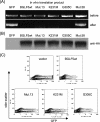
The DNase and shutoff functions of BGLF5 are separable. (A) 293 cells were transfected with pCDNA3-IRES-nlsGFP empty vector or with wild-type (wt) BGLF5 and the different indicated BGLF5 mutant insert vectors. At 48 h after transfection, GFP intensity was analyzed by flow cytometry. (B) Linearized pGEM5Zf(+) DNA was incubated with the indicated BGLF5 mutants synthesized as in vitro translation products. After the degradation reaction, the DNA was resolved by agarose gel electrophoresis and visualized by ethidium bromide staining.
FIG. 6.
FIG. 6.
Inhibition of antigen presentation maps to the shutoff function of BGLF5. (A) 293 cells were cotransfected with 1.0 μg pCEP4-SM plasmid together with 1.0 μg pCDNA3-IRES-nlsGFP empty vector, with wild-type (wt) BGLF5, or with the different indicated BGLF5 mutant vectors. At 24 h posttransfection, the 293 cells were cocultured with effector GLC T-cell clones for a further 18 h, and the supernatants were tested for the release of IFN-γ as for Fig. 2B. (B) Total lysates were generated from the above transfections, and 2 × 10⁵ cell equivalents were separated and analyzed by Western blotting using antibodies specific for BGLF5 or for calregulin, as a loading control. The loading sequence is the same as the sequence shown in panel A.
FIG. 7.
FIG. 7.
(A) Linearized pGEM5Zf(+) DNA was incubated with the indicated HA-tagged BGLF5 mutants synthesized as in vitro translation products. Samples of pGEM5Zf(+) substrate taken before and after the AE degradation reaction were resolved by agarose gel electrophoresis and visualized by ethidium bromide staining. (B) To verify that equal amounts of translated product were added to each enzyme reaction mixture,10 μl of in vitro translation product was separated by SDS-PAGE and analyzed by Western blotting using antibodies specific for the HA tag. (C) Shutoff function was assayed by flow cytometry as for Fig. 5A.

Similar articles

Cited by

References

    1. Babcock, G. J., L. L. Decker, M. Volk, and D. A. Thorley-Lawson. 1998. EBV persistence in memory B cells in vivo. Immunity 9395-404. - PubMed
    1. Barnstable, C. J., W. F. Bodmer, G. Brown, G. Galfre, C. Milstein, A. F. Williams, and A. Ziegler. 1978. Production of a monoclonal antibodies to group A erythrocytes, HLA and other human cell surface antigens: new tools for genetic analysis. Cell 149-20. - PubMed
    1. Buisson, M., E. Manet, M. C. Trescol-Biemont, H. Gruffat, B. Durand, and A. Sergeant. 1989. The Epstein-Barr virus (EBV) early protein EB2 is a posttranscriptional activator expressed under the control of EBV transcription factors EB1 and R. J. Virol. 635276-5284. - PMC - PubMed
    1. Cook, I. D., F. Shanahan, and P. J. Farrell. 1994. Epstein-Barr virus SM protein. Virology 205217-227. - PubMed
    1. Elgadi, M. M., C. E. Hayes, and J. R. Smiley. 1999. The herpes simplex virus vhs protein induces endoribonucleolytic cleavage of target RNAs in cell extracts. J. Virol. 737153-7164. - PMC - PubMed

Publication types

MeSH terms

Substances