. 2008 Mar;82(5):2056-64.
doi: 10.1128/JVI.01803-07. Epub 2007 Dec 19.

Dominant-negative FADD rescues the in vivo fitness of a cytomegalovirus lacking an antiapoptotic viral gene

Affiliations

Dominant-negative FADD rescues the in vivo fitness of a cytomegalovirus lacking an antiapoptotic viral gene

Luka Cicin-Sain et al. J Virol. 2008 Mar.

Abstract

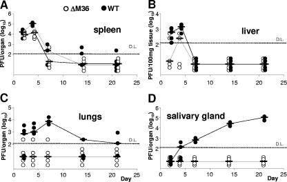
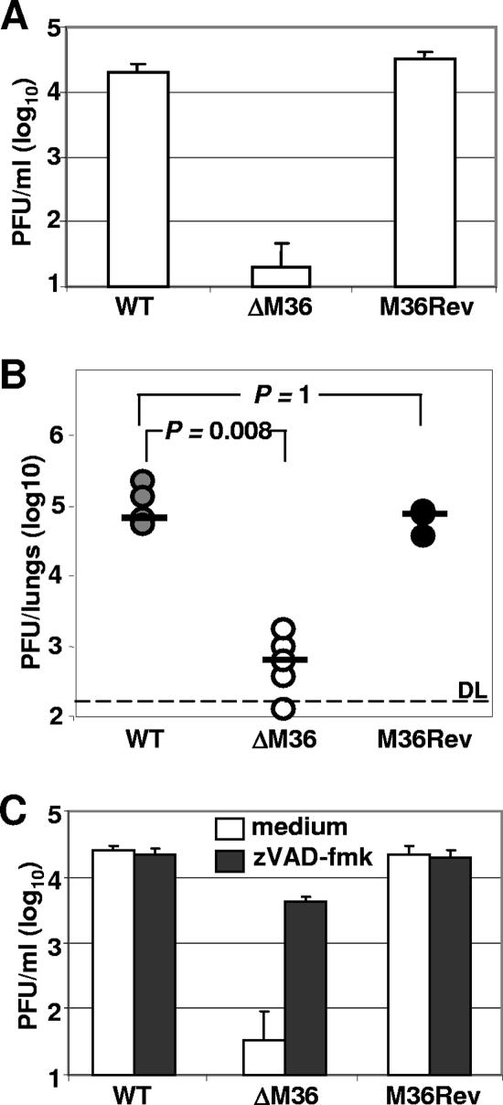
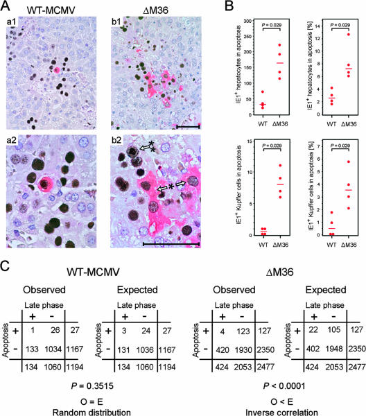
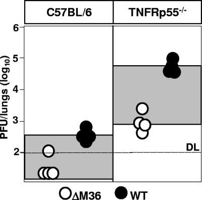
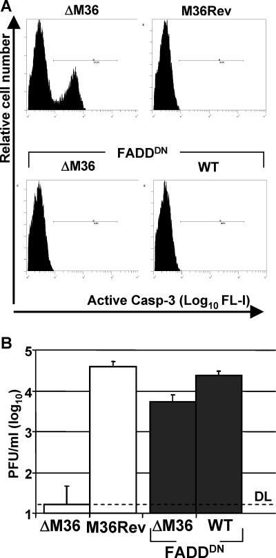
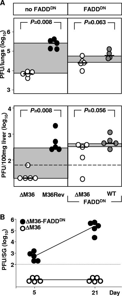
Genes that inhibit apoptosis have been described for many DNA viruses. Herpesviruses often contain even more than one gene to control cell death. Apoptosis inhibition by viral genes is postulated to contribute to viral fitness, although a formal proof is pending. To address this question, we studied the mouse cytomegalovirus (MCMV) protein M36, which binds to caspase-8 and blocks death receptor-induced apoptosis. The growth of MCMV recombinants lacking M36 (DeltaM36) was attenuated in vitro and in vivo. In vitro, caspase inhibition by zVAD-fmk blocked apoptosis in DeltaM36-infected macrophages and rescued the growth of the mutant. In vivo, DeltaM36 infection foci in liver tissue contained significantly more apoptotic hepatocytes and Kupffer cells than did revertant virus foci, and apoptosis occurred during the early phase of virus replication prior to virion assembly. To further delineate the mode of M36 function, we replaced the M36 gene with a dominant-negative FADD (FADD(DN)) in an MCMV recombinant. FADD(DN) was expressed in cells infected with the recombinant and blocked the death-receptor pathway, replacing the antiapoptotic function of M36. Most importantly, FADD(DN) rescued DeltaM36 virus replication, both in vitro and in vivo. These findings have identified the biological role of M36 and define apoptosis inhibition as a key determinant of viral fitness.


Figures

FIG. 1.
FIG. 1.
ΔM36 is attenuated in vivo. BALB/c mice were infected i.p. with 5 × 10⁴ PFU of ΔM36 (○) or WT-MCMV (•). Infectious virus in homogenates of spleen (A), liver (B), lungs (C), and salivary glands (D) was quantitated by plaque assay on days 2, 4, 7, 14, and 21 postinfection. Symbols represent individual mice, and horizontal bars marking the median values are connected. D.L., detection limit of the virus plaque assay.
FIG. 2.
FIG. 2.
M36 is required for MCMV replication in macrophages and in vivo. (A) Comparable growth of M36Rev and WT-MCMV in macrophages. Triplicate cultures of IC-21 macrophages were infected at an MOI of 1 with the viruses indicated. (B) Comparable growth of M36Rev and WT-MCMV in vivo. BALB/c mice were infected i.p. with 5 × 10⁵ PFU of the indicated viruses. Infectious virus in lung homogenates was quantitated by plaque assay on day 5. Circles represent individual mice and horizontal bars mark the median values. DL, detection limit of the virus plaque assay. For group comparisons of interest, P values (Wilcoxon-Mann-Whitney rank sum test, two-tailed) are indicated. Differences are considered as significant and not significant for P < 0.01 and P > 0.05, respectively. (C) Caspase inhibition rescues ΔM36 growth. IC-21 macrophages were infected as described above. Cells were exposed to 30 μM zVAD-fmk (□) or left untreated (□). Infectious virus in SN harvested on day 6 was titrated by plaque assay on MEFs. Histograms indicate mean values; error bars indicate standard deviations.
FIG. 3.
FIG. 3.
Deletion of M36 increases the incidence of early apoptosis of infected hepatocytes in situ. BALB/c mice were immunocompromised by total-body gamma irradiation with a single dose of 7 Gy, and were infected intravenously (i.v.) with either 103 PFU of BAC-cloned WT-MCMV or with 105 PFU of mutant virus ΔM36. The analysis was performed on day 10. (A) Two-color IHC images of liver tissue sections with the intranuclear IE1 protein stained in black as a marker of infection and cytoplasmic active caspase-3 stained in red as a marker of apoptosis. (panels a1, overview; panel a2, detail), infection with WT-MCMV; (panels b1, overview; b2, detail), infection with ΔM36. *, Arrows point to infected and apoptotic IE1+Caspase-3+ hepatocytes. Absence of an intranuclear inclusion body indicates an early stage of infection. **, The arrow points to the nucleus of an infected but nonapoptotic IE1+Caspase-3 hepatocyte. The presence of an intranuclear inclusion body indicates a late phase of infection. Bars, 50 μm. (B) Quantitation of apoptotic IE1+Caspase-3+ hepatocytes (upper panels) and Kupffer cells (lower panels) in absolute terms for a representative tissue section area of 10 mm2 (left panels) and relative to the total number of infected, IE1+ hepatocytes and Kupffer cells, respectively (right panels). Dots represent data from four mice per group with the median value indicated. A P value of <0.05 (Wilcoxon-Mann-Whitney rank sum test, two-tailed) indicates a significant difference. (C) Correlation analysis relating apoptosis to the stage of infection. Infected, IE1+ hepatocytes were counted for a total area of 10 mm2 of liver tissue sections and were classified into four groups according to the expression of active caspase-3 (apoptosis +/−) and the presence of an intranuclear inclusion body (late phase +/−). The data were arranged in 2-by-2 contingency tables (“observed” tables) and compared to the tables expected for a random distribution (“expected” tables). The P values (two-tailed) for random distribution (null hypothesis) were calculated by using the Fisher exact test. The null hypothesis is rejected, and the variables are considered to be inversely correlated if the P value was <0.01 and if the number of double positives observed (O) is less than the number of double positives expected (E).
FIG. 4.
FIG. 4.
ΔM36 growth is not rescued in TNFRp55−/− mice. TNFRp55−/− mice and mice of their parental strain C57BL/6 were i.v. infected with 106 PFU of either ΔM36 (○) or WT-MCMV (•). Virus in lung homogenates was quantified on days 3 by virus plaque assay. Circles represent individual mice. Differences between median values are highlighted by shading. DL, detection limit of the assay.
FIG. 5.
FIG. 5.
Dominant-negative FADD reverses the ΔM36 phenotype in macrophage cell culture. (A) FADDDN inhibits apoptosis in ΔM36-infected macrophages. IC-21 macrophages were infected with the indicated viruses at an MOI of 1. At 24 h after infection, apoptotic macrophages were quantitated by cytofluorometric analysis using the expression of active caspase-3 (abscissa) as a marker of apoptosis. FL-I, fluorescence intensity (B) FADDDN rescues the growth of ΔM36 in macrophages. IC-21 macrophages were infected at an MOI of 1 with the indicated viruses devoid of FADDDN (□) or encoding FADDDN (▪). Virus in SN was quantitated on day 5 after infection. Histograms represent mean values from three independent experiments. Error bars indicate standard deviations. DL, detection limit of the virus plaque assay.
FIG. 6.
FIG. 6.
Dominant-negative FADD reverses the ΔM36 phenotype in host organs. (A) BALB/c mice were infected i.v. with 3 × 10⁵ PFU of MCMV expressing FADDDN (ΔM36-FADDDN or WT-FADDDN) and, for a control, with the corresponding viruses not encoding FADDDN. The absence of M36 is symbolized by open circles, and the presence of M36 in M36Rev and WT-MCMV (WT) is symbolized by black-filled and gray-filled circles, respectively. The amount of infectious virus in the lungs (upper panel) and liver (lower panel) was determined on day 5 after infection. Circles represent data from individual mice. Differences in median values are highlighted by gray shading. The dashed line indicates the detection limit of the virus plaque assay for liver homogenate. For group comparisons of interest, the P values (Wilcoxon-Mann-Whitney rank sum test, two-tailed) are indicated. Differences are considered significant and not significant for P < 0.01 and P > 0.05, respectively. (B) BALB/c mice were infected i.p. with 5 × 10⁵ PFU of ΔM36 (○) or ΔM36-FADDDN (•). Virus titers are represented on the y axis; times after infection are represented on the x axis. Infectious virus in salivary glands (SG) was determined on days 5 and 21 after infection. Circles represent data from individual mice. Median values are connected with solid lines.

References

    1. Altmann, M., and W. Hammerschmidt. 2005. Epstein-Barr virus provides a new paradigm: a requirement for the immediate inhibition of apoptosis. PLoS Biol. 3e404. - PMC - PubMed
    1. Andoniou, C. E., and M. A. Degli-Esposti. 2006. Insights into the mechanisms of CMV-mediated interference with cellular apoptosis. Immunol. Cell Biol. 8499-106. - PubMed
    1. Askenasy, N., E. S. Yolcu, I. Yaniv, and H. Shirwan. 2005. Induction of tolerance using Fas ligand: a double-edged immunomodulator. Blood 1051396-1404. - PubMed
    1. Bertin, J., R. C. Armstrong, S. Ottilie, D. A. Martin, Y. Wang, S. Banks, G. H. Wang, T. G. Senkevich, E. S. Alnemri, B. Moss, M. J. Lenardo, K. J. Tomaselli, and J. I. Cohen. 1997. Death effector domain-containing herpesvirus and poxvirus proteins inhibit both Fas- and TNFR1-induced apoptosis. Proc. Natl. Acad. Sci. USA 941172-1176. - PMC - PubMed
    1. Brune, W., C. Menard, J. Heesemann, and U. H. Koszinowski. 2001. A ribonucleotide reductase homolog of cytomegalovirus and endothelial cell tropism. Science 291303-305. - PubMed

Publication types

LinkOut - more resources