Comparative Study
. 2007 Dec 19;27(51):13982-90.
doi: 10.1523/JNEUROSCI.4226-07.2007.

The G59S mutation in p150(glued) causes dysfunction of dynactin in mice

Affiliations
Comparative Study

The G59S mutation in p150(glued) causes dysfunction of dynactin in mice

Chen Lai et al. J Neurosci. .

Abstract

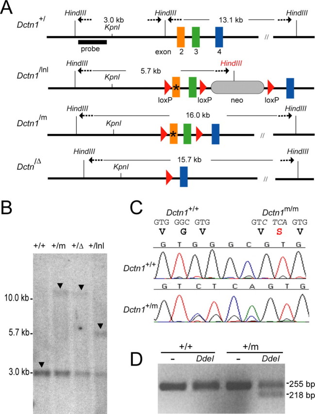
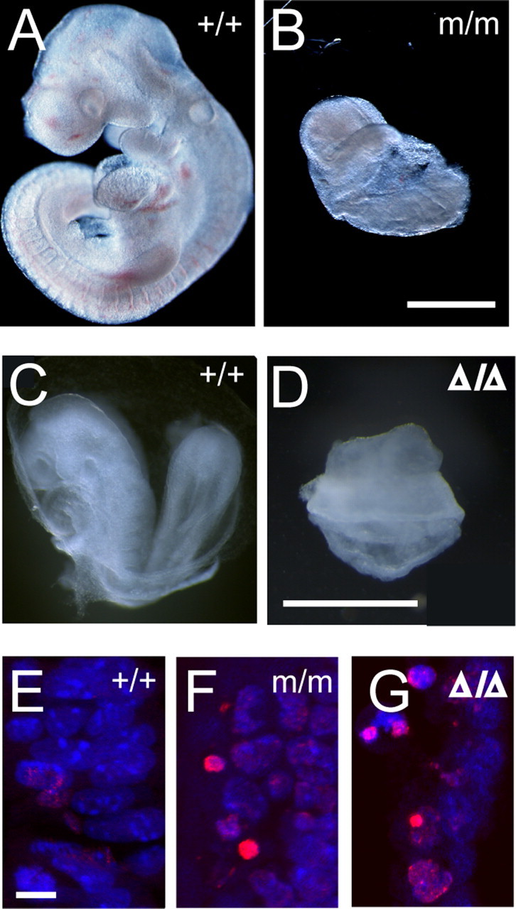
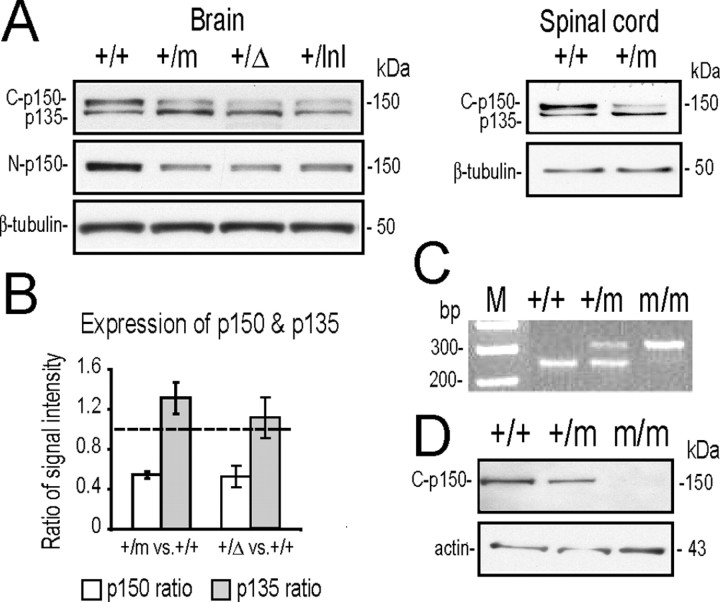
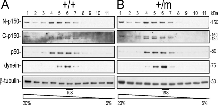
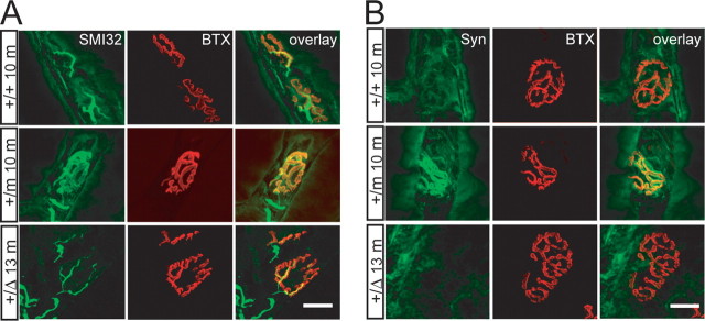
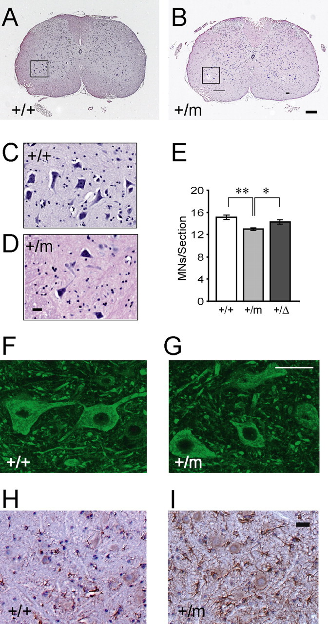
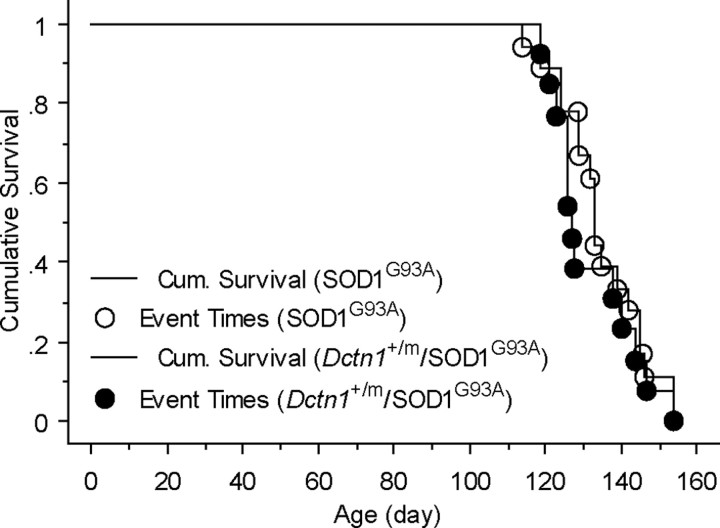
The G59S missense mutation at the conserved microtubule-binding domain of p150(glued), a major component of dynein/dynactin complex, has been linked to an autosomal dominant form of motor neuron disease (MND). To study how this mutation affects the function of the dynein/dynactin complex and contributes to motor neuron degeneration, we generated p150(glued) G59S knock-in mice. We found that the G59S mutation destabilizes p150(glued) and disrupts the function of dynein/dynactin complex, resulting in early embryonic lethality of homozygous knock-in mice. Heterozygous knock-in mice, which developed normally, displayed MND-like phenotypes after 10 months of age, including excessive accumulation of cytoskeletal and synaptic vesicle proteins at neuromuscular junctions, loss of spinal motor neurons, increase of reactive astrogliosis, and shortening of gait compared with wild-type littermates and age-matched p150(glued) heterozygous knock-out mice. Our findings indicate that the G59S mutation in p150(glued) abrogates the normal function of p150(glued) and accelerates motor neuron degeneration.


Conflict of interest statement

The authors declare no competing financial interests.

Figures

Figure 1.
Figure 1.
Generation of p150glued G59S knock-in mice. A, A schematic outline of mouse Dctn1 wild-type (+), floxed neomycin gene (lnl) insertion, G59S knock-in mutant (m), and exons 2 and 3 deletion (Δ) alleles. * represents the mutated region. B, Southern blot analysis of genomic DNA extracted from wild-type and heterozygous mutant mice confirmed the correct targeting of mutated Dctn1 alleles, which displayed 16.0, 15.7, 5.7, and 3.0 kb HindIII fragments (arrows) for the lnl, m, Δ, and + alleles, respectively. C, Chromatograms showed the partial sequence of exon2 amplified from RNA extracted from Dctn1 wild-type (+/+) and heterozygous knock-in (+/m) mice. D, Agarose gel electrophoresis of the RT-PCR products from wild-type (+/+) and Dctn1 +/m (+/m) after digestion with (+) or without (−) DdeI.
Figure 2.
Figure 2.
Homozygous Dctn1 knock-in mice were embryonic lethal. A, B, Wild-type (+/+) and Dctn1 m/m (m/m) embryos at 9.5 dpc. Scale bar, 500 μm. C, D, Wild-type (+/+) and Dctn1 Δ/Δ (Δ/Δ) were at 8.5 dpc. Scale bar, 600 μm. E–G, TUNEL staining (red) revealed increased programmed cell death in Dctn1 m/m (m/m; F) and Dctn1 Δ/Δ (Δ/Δ; G) embryos compared with wild-type controls (+/+; E). Nuclei were stained with ToPro-3 (blue). Scale bar, 10 μm.
Figure 3.
Figure 3.
The G59S mutation destabilized p150glued protein. A, Western blot analyses revealed the expression of p150glued/p135 (top; detected by the p150glued C-terminal antibody, C-p150) and p150glued alone (middle; detected by p150glued N-terminal antibody, N-p150) in brains and spinal cords of wild-type (+/+), Dctn1 +/m (+/m), Dctn1 +/Δ (+/Δ), and Dctn1 +/lnl (+/lnl) mice. The expression of β-tubulin (bottom) was used as a loading control. B, Bar graph shows reduced accumulation of p150glued in Dctn1 +/m and Dctn1 +/Δ brains compared with wild-type controls, whereas the expression of alternative spliced p135 was slightly increased in these animals. Data are means ± SEM. C, Genotyping of wild-type (+/+), Dctn1 +/m (+/m), and Dctn1 m/m (m/m) embryos by PCR amplification of genomic DNA prepared from yolk sacs. D, Western blot analyses using p150glued C-terminal antibody revealed the absence of p150glued protein from Dctn1 m/m (m/m) embryos (top). The expression of β-actin (bottom) was used as a loading control.
Figure 4.
Figure 4.
Dynein/dynactin complex remains intact in Dctn1 +/m mice. Density gradient centrifugation showed that p150glued and its alternative splicing variant, p135, predominantly migrated at ∼19S in a 5–20% sucrose gradient similar to dynein and p50 in wild-type (+/+) (A) and Dctn1 +/m (+/m) (B) samples prepared from brains. The expression of β-tubulin was used here as a loading control.
Figure 5.
Figure 5.
Accumulation of neurofilament and synaptophysin at the NMJ of Dctn1 +/m mice. A, Motor neurons axons revealed by SMI32 staining terminated at endplates visualized by BTX staining. Increased accumulation of NF was observed at NMJ of gastrocnemius muscle sections from Dctn1 +/m (+/m 10m) mice at 10 months of age compared with wild-type (+/+ 10 m) littermate controls and Dctn1 +/Δ (+/Δ 13 m) mice at 13 months of age. B, Similar to NF, elevated accumulation of synaptophysin (Syn) was observed at NMJ of gastrocnemius muscle sections from Dctn1 +/m (+/m 10 m) mice at 10 months of age compared with wild-type (+/+ 10 m) littermate controls and Dcrn1 +/Δ (+/Δ 13 m) mice at 13 months of age. Muscle section thickness, 10 μm. Scale bars, 20 μm.
Figure 6.
Figure 6.
Motor neuron degeneration in Dctn1 +/m mice (A–D) HE staining of coronal sections of lumbar spinal cords revealed motor neurons in wild-type (A, C) and Dctn1 +/m (B, D) mice at 16 months of age. The insets (C, D) showed motor neurons under higher magnification of the boxed area (A, B). Scale bars: main, 100 μm; insets, 50 μm. E, Box graph of numbers of motor neurons per lumbar spinal cord section of wild-type (+/+), Dctn1 +/m (+/m), and Dctn1 +/Δ (+/Δ) mice. *p < 0.01 and **p < 0.001, respectively. F, G, Representative images of p150glued staining with the antibody against the C-terminal of p150glued in the wild-type (+/+) and Dctn1 +/m (+/m) lumbar spinal cords. Scale bar, 50 μm. H, I, Representative images of GFAP staining in the wild-type (+/+) and Dctn1 +/m (+/m) lumbar spinal cords. All the sections were counterstained with HE. Scale bar, 20 μm.
Figure 7.
Figure 7.
The G59S mutation in p150glued does not affect the survival of SOD1G93A transgenic mice. Kaplan–Meier plot of cumulative probability of survival of SOD1G93A/Dctn1 +/+ (n = 10) and SOD1G93A/Dctn1 +/m mice (n = 15).

References

    1. Allan V. Motor proteins: a dynamic duo. Curr Biol. 1996;6:630–633. - PubMed
    1. Bross P, Corydon TJ, Andresen BS, Jorgensen MM, Bolund L, Gregersen N. Protein misfolding and degradation in genetic diseases. Hum Mutat. 1999;14:186–198. - PubMed
    1. Culver-Hanlon TL, Lex SA, Stephens AD, Quintyne NJ, King SJ. A microtubule-binding domain in dynactin increases dynein processivity by skating along microtubules. Nat Cell Biol. 2006;8:264–270. - PubMed
    1. Eaton BA, Fetter RD, Davis GW. Dynactin is necessary for synapse stabilization. Neuron. 2002;34:729–741. - PubMed
    1. Glass JD, Griffin JW. Neurofilament redistribution in transected nerves: evidence for bidirectional transport of neurofilaments. J Neurosci. 1991;11:3146–3154. - PMC - PubMed

Publication types