Comparative Study
. 2007 Dec 19;27(51):14007-11.
doi: 10.1523/JNEUROSCI.3587-07.2007.

Synaptic strength of individual spines correlates with bound Ca2+-calmodulin-dependent kinase II

Affiliations
Comparative Study

Synaptic strength of individual spines correlates with bound Ca2+-calmodulin-dependent kinase II

Brent Asrican et al. J Neurosci. .

Abstract

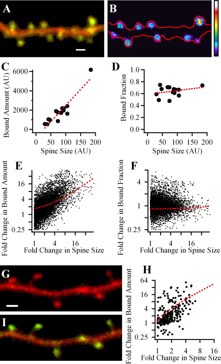
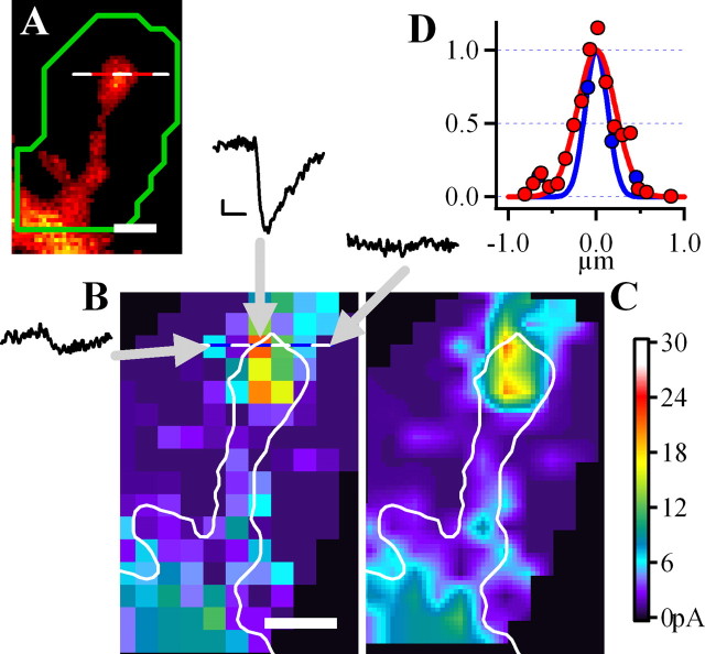
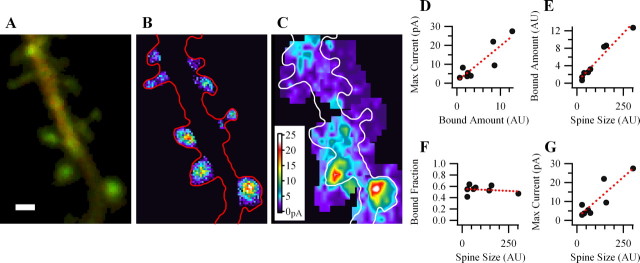
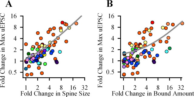
Both synaptic strength and spine size vary from spine to spine, but are strongly correlated. This gradation is regulated by activity and may underlie information storage. Ca2+-calmodulin-dependent kinase II (CaMKII) is critically involved in the regulation of synaptic strength and spine size. The high amount of the kinase in the postsynaptic density has suggested that the kinase has a structural role at synapses. We demonstrated previously that the bound amount of CaMKIIalpha in spines persistently increases after induction of long-term potentiation, prompting the hypothesis that this amount may correlate with synaptic strength. To test this hypothesis we combined two recently developed methods, two-photon uncaging of glutamate for determining the EPSC of individual spines (uEPSC) and quantitative microscopy for measuring bound CaMKIIalpha in the same spines. We found that under basal conditions the relative bound amount of CaMKIIalpha varied over a 10-fold range and positively correlated with the uEPSC. Both the bound amount of CaMKIIalpha in spines and uEPSC also positively correlated with spine size. Interestingly, the bound CaMKIIalpha fraction (bound/total CaMKIIalpha in spines) remained remarkably constant across all spines. The results are consistent with the hypothesis that bound CaMKII serves as a structural organizer of postsynaptic molecules and thereby may be involved in maintaining spine size and synaptic strength.


Figures

Figure 1.
Figure 1.
Bound amount of GFP-CaMKIIα strongly correlates with spine volume but bound fraction does not. A, Merged image of a dendritic segment of neuron expressing mGFP-CaMKIIα and infused with Alexa 594 dye. B, A map of the bound mGFP-CaMKIIα in spines in pseudocolor coding; warmer colors correspond to larger bound amount. Red outline shows the dendrite morphology. BA was calculated only in clearly identified spines. Scale, 1 μm. C, D, Plots of data obtained from the dendrite shown in A and B. Each symbol shows the value for an individual spine. Dashed line, Linear fit of data. E, F, Summary data for the correlations of spine BA or BF of GFP-CaMKIIα and spine volume. Each data point is a result of comparison of two spines from the same dendrite. E, Fold change in BA versus fold change in spine size. F, Fold change in BF versus fold change in spine size. G, H, Measuring BA using PAGFP-CaMKIIα. Merged image of a dendritic segment of neuron expressing PAGFP-CaMKIIα and mRFP(Cherry) before (G) and 2 min after (I) the photoactivation of PAGFP. H, Summary graph, showing fold change in spine size versus fold change in BA of PAGFP-CaMKIIα, reveals strong correlation between these parameters.
Figure 2.
Figure 2.
Measuring the postsynaptic strength of a single spine using glutamate uncaging. A, A spine from a neuron filled with Alexa 594 dye with ROI for uncaging shown in green; scale, 1 μm. B, The map of uncaging locations within the ROI indicated in A. Three examples of uEPSCs are from locations indicated by arrows. Calibration: 5pA, 5 ms. Different uncaging locations yielded uEPSCs ranging from negligible to 25 pA. C, The same map but smoothed by interpolation. D, The spatial distribution of fluorescence along the dotted line in A (red) showing that the spine diameter as estimated by full width at half maximum (FWHM) is ∼0.5 μm. Blue line, The distribution of the uncaging response, which is slightly narrower (FWHM, 0.4 μm) than the diameter of the spine.
Figure 3.
Figure 3.
At individual spines, the maximal uEPSC positively correlates with the relative bound amount of GFP-CaMKIIα. A, An overlay of green and red fluorescence from a dendrite expressing GFP-CaMKIIα (green) and mRFP (red). B, Calculated locations of relative bound GFP-CaMKIIα amount. C, uEPSCs maps for spines of the same dendrite. D–G, Plots showing dependence of maximal postsynaptic uEPSC on the amount of bound CaMKII and dependence of CaMKII bound amount, bound fraction and uEPSC on spine volume for the spines shown in A. Scale bar, 1 μm.
Figure 4.
Figure 4.
Summary data for the correlation of postsynaptic uEPSC with spine bound amount of GFP-CaMKIIα or spine volume. Each data point is a result of comparison of two spines from the same dendrite. A, B, Each graph shows a fold change in one parameter versus fold change of another parameters: uEPSC versus spine volume (A) or versus bound amount of CaMKII (B). In A and B, data for each dendrite is color coded. Gray lines, Linear fits.

References

    1. Allison DW, Chervin AS, Gelfand VI, Craig AM. Postsynaptic scaffolds of excitatory and inhibitory synapses in hippocampal neurons: maintenance of core components independent of actin filaments and microtubules. J Neurosci. 2000;20:4545–4554. - PMC - PubMed
    1. Asrican B, De Koninck P, Lisman J, Otmakhov N. Measuring CaMKII binding to different postsynaptic targets in live hippocampal neurons. Soc Neurosci Abstr. 2006;32:633–638.
    1. Barria A, Malinow R. NMDA Receptor Subunit Composition Controls Synaptic Plasticity by Regulating Binding to CaMKII. Neuron. 2005;48:289–301. - PubMed
    1. Cheng D, Hoogenraad CC, Rush J, Ramm E, Schlager MA, Duong DM, Xu P, Wijayawardana SR, Hanfelt J, Nakagawa T, Sheng M, Peng J. Relative and absolute quantification of postsynaptic density proteome isolated from rat forebrain and cerebellum. Mol Cell Proteomics. 2006;5:1158–1170. - PubMed
    1. Colbran RJ. Targeting of calcium/calmodulin-dependent protein kinase II. Biochem J. 2004;378:1–16. - PMC - PubMed

Publication types

MeSH terms

Substances

LinkOut - more resources