Comparative Study
. 2007 Dec 19;27(51):14041-8.
doi: 10.1523/JNEUROSCI.3217-07.2007.

Effects of saporin-induced lesions of three arousal populations on daily levels of sleep and wake

Affiliations
Comparative Study

Effects of saporin-induced lesions of three arousal populations on daily levels of sleep and wake

Carlos Blanco-Centurion et al. J Neurosci. .

Abstract

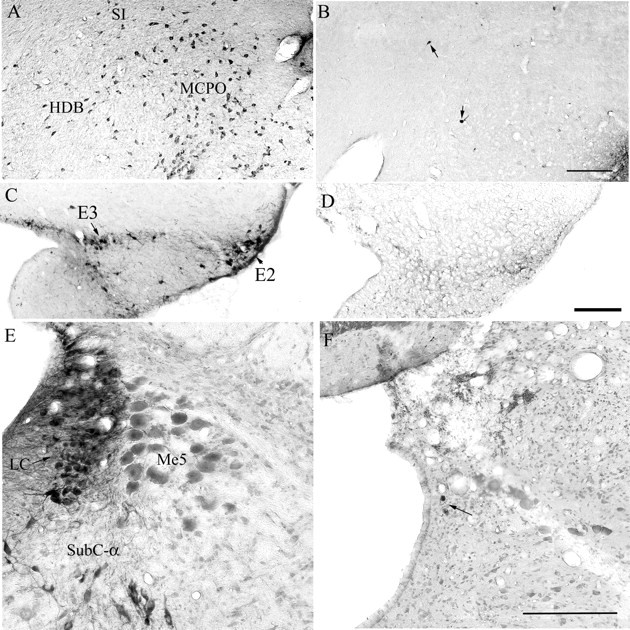
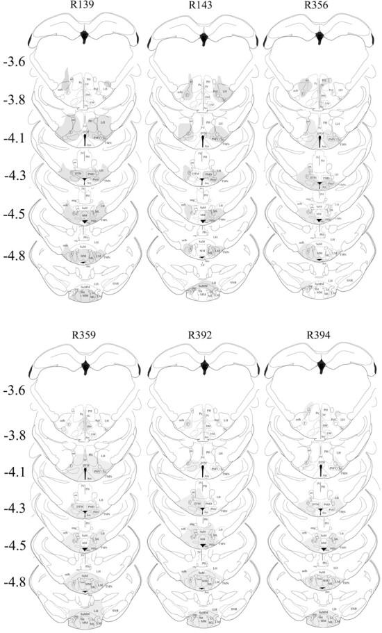
The hypocretin (HCRT) neurons are located only in the perifornical area of the lateral hypothalamus and heavily innervate the cholinergic neurons in the basal forebrain (BF), histamine neurons in the tuberomammillary nucleus (TMN), and the noradrenergic locus ceruleus (LC) neurons, three neuronal populations that have traditionally been implicated in regulating arousal. Based on the innervation, HCRT neurons may regulate arousal by driving these downstream arousal neurons. Here, we directly test this hypothesis by a simultaneous triple lesion of these neurons using three saporin-conjugated neurotoxins. Three weeks after lesion, the daily levels of wake were not changed in rats with double or triple lesions, although rats with triple lesions were asleep more during the light-to-dark transition period. The double- and triple-lesioned rats also had more stable sleep architecture compared with nonlesioned saline rats. These results suggest that the cholinergic BF, TMN, and LC neurons jointly modulate arousal at a specific circadian time, but they are not essential links in the circuitry responsible for daily levels of wake, as traditionally hypothesized.


Figures

Figure 1.
Figure 1.
The loss of neurons in three arousal populations after administration of three saporin-conjugated neurotoxins. Photomicrographs A, C, and E depict the cholinergic neurons in the BF, the histamine neurons of the TMN, and the noradrenergic neurons of the LC in saline-treated rats. The cholinergic BF neurons were destroyed with intracerebroventricular injection of 192-IgG-saporin (photo B). The TMN neurons were destroyed with hypocretin-2-saporin (photo D) and the noradrenergic LC neurons were destroyed with anti-dopamine-β-hydroxylase-saporin (photo F). Scale bars: B, D, F, 200 μm. The arrows point to few surviving neurons. E2–E3, Histaminergic clusters within the TMN according to Inagaki et al. (1990); HDB, horizontal limb of the diagonal band of Broca; MCPO, magnocellular preoptic nucleus; Me5, mesencephalic trigeminal nucleus; SI, substantia innominata; SubC-α, subceruleus α.
Figure 2.
Figure 2.
Extent of neuronal loss because of local injection of HCRT2-SAP (250 ng/μl in 0.25 μl each side) into the TMN. HCRT2-SAP kills neurons that possess the HCRT-2 receptor, which is on the histaminergic TMN neurons but also on other adjacent neurons. Thus, the neuronal marker NeuN was used to assess the extent of nonspecific neuronal loss, and those regions devoid of neurons were then drawn onto the corresponding figures taken from the rat brain atlas (Paxinos and Watson, 2006). Each column represents an individual rat (marked by number at the top of each column) in the triple-lesion group (BF+TMN+LC). The numbers to the left indicate anteroposterior distance from bregma in millimeters. Arc, Arcuate n; DM, dorsomedial hypothalamus; DTM, dorsal tuberomammillary n; MM, medial mammillary n; MMn, median mammillary n; ML, medial mammillary n lateral; LH, lateral hypothalamus; LM, lateral mammillary n; PeF, perifornical area; PH, posterior hypothalamus; PMD, premammillary n dorsal; SuM, supramammillary n; SNR, substantia nigra reticular; VM, ventromedial hypothalamus; Te, Terete hypothalamic n; 3V, third ventricle; f, fornix; mfb, medial forebrain bundle; mt, mammilothalamic tract; mtg, mammilotegmental tract; mp, mammillary peduncle.
Figure 3.
Figure 3.
Percentage (±SEM) of wake, non-REM, and REM sleep in lesioned and nonlesioned rats. The line graphs summarize the average in 4 h blocks over the 24 h period with the data double-plotted to illustrate the diurnal rhythm of the sleep–wake cycle. The crescent moon symbols represent the 12 h dark cycle. The bar graphs on the right summarize the data during the 12 h day and night cycles. *p < 0.01 versus triple-lesioned rats (post hoc significance after one-way ANOVA); **p < 0.05 versus double- and triple-lesioned rats (post hoc after one-way ANOVA); formula image
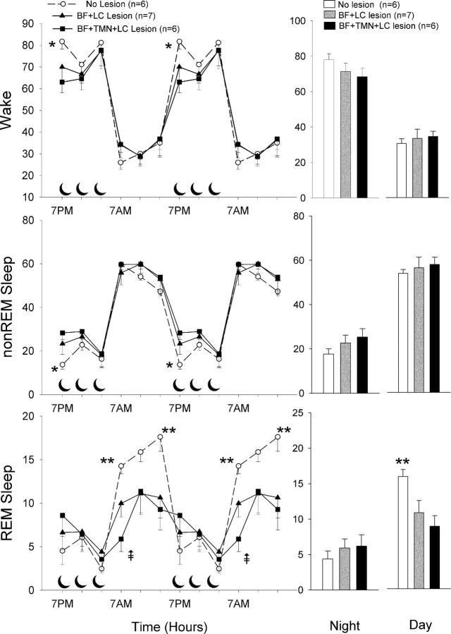
p < 0.05 versus double lesion (post hoc after one-way ANOVA).
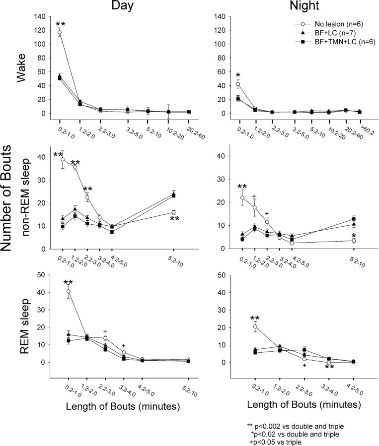
Figure 4.
Figure 4.
The number and length of bouts of wake, non-REM, and REM, sleep in saline versus lesioned rats. Each bout of wake, non-REM, and REM sleep was placed into a bin corresponding to a specific length (in minutes) of the bout, and the figure represents the average (±SEM) number of bouts of a specific length for the lesion and saline groups. *p < 0.02 saline versus double and triple; **p < 0.002 saline versus double- and triple-lesioned rats; +p < 0.05 saline versus triple-lesioned rats. Significance represents post hoc test after one-way ANOVA.

References

    1. Beuckmann CT, Sinton CM, Williams SC, Richardson JA, Hammer RE, Sakurai T, Yanagisawa M. Expression of a poly-glutamine-ataxin-3 transgene in orexin neurons induces narcolepsy-cataplexy in the rat. J Neurosci. 2004;24:4469–4477. - PMC - PubMed
    1. Blanco-Centurion C, Gerashchenko D, Salin-Pascual RJ, Shiromani PJ. Effects of hypocretin2-saporin and antidopamine-beta-hydroxylase-saporin neurotoxic lesions of the dorsolateral pons on sleep and muscle tone. Eur J Neurosci. 2004;19:2741–2752. - PMC - PubMed
    1. Blanco-Centurion C, Xu M, Murillo-Rodriguez E, Gerashchenko D, Shiromani AM, Salin-Pascual RJ, Hof PR, Shiromani PJ. Adenosine and sleep homeostasis in the basal forebrain. J Neurosci. 2006a;26:8092–8100. - PMC - PubMed
    1. Blanco-Centurion CA, Shiromani A, Winston E, Shiromani PJ. Effects of hypocretin-1 in 192-IgG-saporin-lesioned rats. Eur J Neurosci. 2006b;24:2084–2088. - PubMed
    1. Bourgin P, Huitron-Resendiz S, Spier AD, Fabre V, Morte B, Criado JR, Sutcliffe JG, Henriksen SJ, de Lecea L. Hypocretin-1 modulates rapid eye movement sleep through activation of locus coeruleus neurons. J Neurosci. 2000;20:7760–7765. - PMC - PubMed

Publication types

LinkOut - more resources