Comparative Study
. 2007 Dec 19;27(51):14117-27.
doi: 10.1523/JNEUROSCI.3884-07.2007.

Expression and localization of RGS9-2/G 5/R7BP complex in vivo is set by dynamic control of its constitutive degradation by cellular cysteine proteases

Affiliations
Comparative Study

Expression and localization of RGS9-2/G 5/R7BP complex in vivo is set by dynamic control of its constitutive degradation by cellular cysteine proteases

Garret R Anderson et al. J Neurosci. .

Abstract

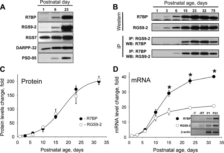
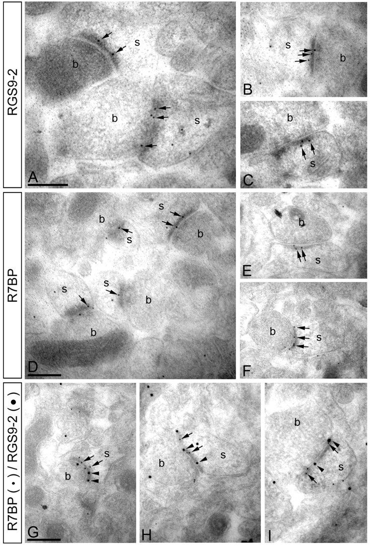
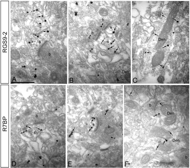
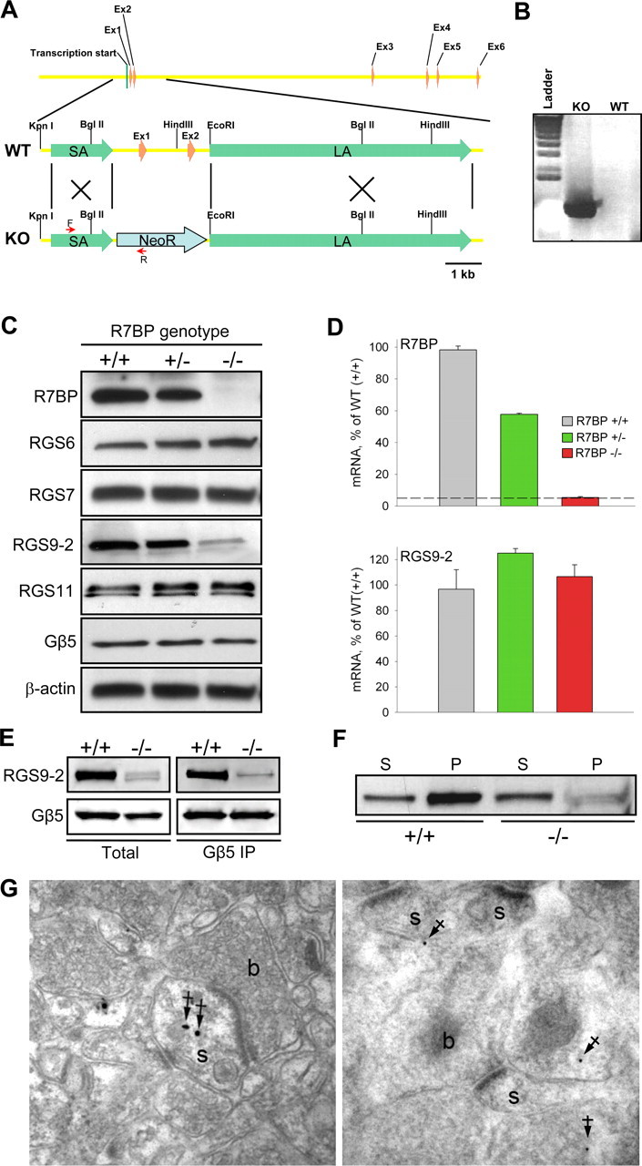
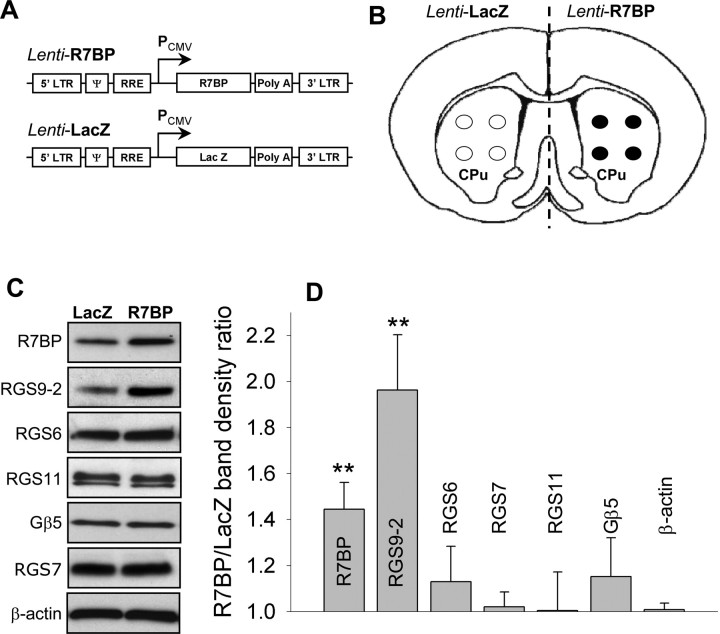
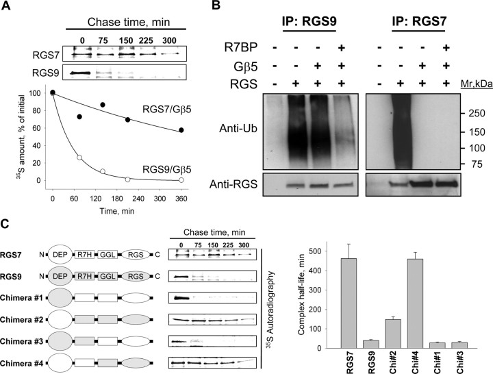
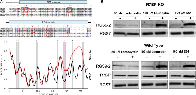
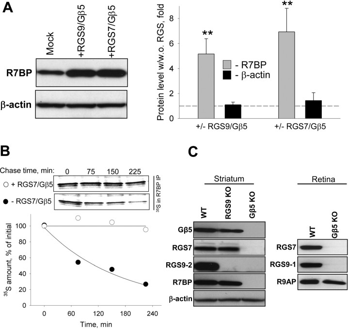
A member of regulator of G-protein signaling family, RGS9-2, is an essential modulator of signaling through neuronal dopamine and opioid G-protein-coupled receptors. Recent findings indicate that the abundance of RGS9-2 determines sensitivity of signaling in the locomotor and reward systems in the striatum. In this study we report the mechanism that sets the concentration of RGS9-2 in vivo, thus controlling G-protein signaling sensitivity in the region. We found that RGS9-2 possesses specific degradation determinants which target it for constitutive destruction by lysosomal cysteine proteases. Shielding of these determinants by the binding partner R7 binding-protein (R7BP) controls RGS9-2 expression at the posttranslational level. In addition, binding to R7BP in neurons targets RGS9-2 to the specific intracellular compartment, the postsynaptic density. Implementation of this mechanism throughout ontogenetic development ensures expression of RGS9-2/type 5 G-protein beta subunit/R7BP complexes at postsynaptic sites in unison with increased signaling demands at mature synapses.


Figures

Figure 1.
Figure 1.
Coregulation of RGS9-2 and R7BP expression during differentiation of striatal neurons. A, Western blot analysis of change in protein expression level on postnatal differentiation of striatum in Swiss-Webster mice. Striatal lysates (20 μg/lane) were analyzed for protein expression using specific antibodies. B, Time course of RGS9-2 and R7BP expression and their complex formation at the protein level. RGS9-2 and R7BP were immunoprecipitated (IP) and subjected to Western blot (WB) analysis. Immunoprecipitation efficiency in the experiments was >90%. C, Quantification of changes in levels of proteins from three separate groups analyzed as described in B. Band densities were determined by NIH ImageJ software and used to determine changes in protein levels relative to day 1 as described in Materials and Methods. D, Quantitative analysis of R7BP and RGS9-2 expression in development at mRNA level. Relative levels of RGS9-2 and R7BP mRNAs were measured by quantitative RT-PCR and normalized to the levels of β-actin mRNA amplified in parallel as an endogenous reference. Relative quantification algorithm was used in which changes in amplification threshold were normalized to sample from postnatal day 1. Data are averaged from three separate groups of animals. Inset shows gel electrophoresis of PCR products amplified from newborn (P1) or adult (P23) samples. No reverse transcriptase (-RT) or template (-T) was added in controls. *p < 0.05, statistically significant differences between relative changes in R7BP and RGS9-2 mRNA (t test).
Figure 2.
Figure 2.
RGS9-2 and R7BP colocalize at postsynaptic densities of excitatory synapses. Electron micrographs of the striatum showing immunoreactivity for RGS9-2 and R7BP as detected using a postembedding immunogold method. A–C, Immunoparticles for RGS9-2 were found within the synaptic specialization of asymmetrical synapses (arrowheads). Gold particles were also observed at the extrasynaptic plasma membrane of spines (s) and to a lesser extent at presynaptic sites along the plasma membrane of axon terminals (b). D–F, Similarly, immunoparticles for R7BP were found within the synaptic specialization of asymmetrical synapses (arrowheads). A few immunoparticles were also observed at the extrasynaptic plasma membrane of spines (s) and at presynaptic sites along the plasma membrane of axon terminals (b). G–I, Double immunogold labeling showing colocalization of RGS9-2 (20 nm particles, arrowheads) and R7BP (10 nm particles, arrows) in individual synapses on spines (s) in the striatum. Scale bars, 0.2 μm.
Figure 3.
Figure 3.
RGS9-2 and R7BP share the same subcellular localization in striatum. Electron micrographs of the striatum showing immunoreactivity for RGS9-2 (A–C) and R7BP (D–F), as detected using a preembedding immunogold method. A–C, Immunoparticles for RGS9-2 were detected associated or close (arrows) to the extrasynaptic plasma membrane of dendritic shafts (Den) and dendritic spines (s) establishing excitatory asynapses with axon terminals (b). Some immunoparticles for RGS9-2 were also detected intracellularly (crossed arrows) associated with intracellular membranes of dendritic shafts and spines. To a lesser extent, RGS9-2 immunoparticles were detected at presynaptic sites associated with the plasma membrane of axon terminals (b) (arrowheads). D–F, Immunoparticles for R7BP were detected associated or close (arrows) to the extrasynaptic plasma membrane of dendritic shafts (Den) and dendritic spines (s) establishing excitatory asynapses with axon terminals (b). Immunoparticles for R7BP were also detected intracellularly (crossed arrows) associated with intracellular membranes of dendritic shafts and spines. A few immunoparticles for R7BP were also detected at presynaptic sites associated with the plasma membrane of axon terminals (b) (arrowheads).
Figure 4.
Figure 4.
Knock-out of R7BP selectively destabilizes RGS9-2 at posttranscriptional level. A, R7BP gene structure and strategy for targeting homologous recombination. Targeting construct contained neomycin resistance cassette (NeoR) flanked by short (SA) and long (LA) homology arms. The positions of primers used to confirm the replacement are shown as red arrows. B, Confirmation of homologous recombination in genomic DNA isolated from the founder animal (KO) compared with wild-type parental strain (WT) by PCR. C, Western blot analysis of protein expression in the brains of wild-type (+/+), heterozygous (+/−) and R7BP knock-out (−/−) mice. The blot shows a representative experiment of three conducted. D, Analysis of R7BP and RGS9-2 transcript levels by quantitative RT-PCR. Total mRNA was isolated from striatal regions of adult mice was quantified by UV spectroscopy, and its equal amounts were subjected to Q-RT PCR amplification. Each experiment was conducted with samples isolated from two to three mice. Dashed line indicates the limiting level for mRNA detection. Error bars represent SEM. E, Immunoprecipitation of RGS9-2/Gβ5 complexes from wild-type and R7BP knock-out striatal tissues. Extraction and immunoprecipitation with anti-Gβ5 antibody was conducted as described in Materials and Methods. Two milligrams of total protein were used in each immunoprecipitation experiment with 14 μg of anti-Gβ5 antibody. Equal protein amounts of whole-cell extracts and volumes of the immunoprecipitation eluates were loaded in each well. The data are representative of three experiments conducted. F, Subcellular fractionation of striatal neurons into synaptic and plasma membrane containing pellet (P) and supernatant (S) containing primarily cytosol and small microsomal membranes. Striatal regions of wild-type (WT) and R7BP knock-out (R7BP KO) mice were homogenized and subjected to one step centrifugation as described in Materials and Methods. Knock-out samples were loaded at four times excess of total protein over wild-type samples. G, Preembedding (left) and postembedding (right) immunogold EM analysis of RGS9-2 localization in striatum of R7BP knock-out mice. Filled arrows indicate location of immunogold particles at intracellular sites, a predominant localization pattern of RGS9-2 in neurons of R7BP knock-outs. s, Dendritic spines; b, plasma membrane of the axon terminals.
Figure 5.
Figure 5.
Lentivirus-mediated overexpression of R7BP in the striatum. A, Schematic representation of lentiviral constructs used for the expression experiments. Open reading frames for R7BP or LacZ were placed under the control of strong cytomegalovirus promoter in the context of lentiviral packaging elements (LTR, Ψ, and RRE). B, Strategy for the lentivirus mediated protein expression in vivo. Each side of the brain received four injections with either R7BP or control LacZ lentiviruses within the caudate/putamen region (CPu). C, Western blot analysis of protein expression in the CPu induced after lentiviral transduction. D, Quantification of Western blot data shown in C from three independent experiments. Band densities were determined by densitometry and used to calculate the ratio of band intensity between R7BP side to LacZ side. **p < 0.01, statistically significant changes in the levels of proteins compared with changes in β-actin expression (last bar) (t test). Error bars are SEM.
Figure 6.
Figure 6.
Selective degradation of RGS9-2/Gβ5 in the absence of R7BP is determined by N-terminal instability elements. A, Pulse-chase metabolic labeling experiments comparing degradation rates of RGS7 and RGS9-1 in 293FT cells. The blot shows the time course of 35S-Met/Cys dissipation from immunoprecipitated RGS proteins, quantified on a graph below. Approximately 25% of recombinant proteins was recovered in the immunoprecipitation eluates. B, Detection of RGS protein modification with ubiquitin in transfected 293FT cells. Top, Ubiquitination of RGS7 or RGS9-2 in the presence or absence of Gβ5 and R7BP was detected by Western blotting with anti-ubiquitin antibodies after protein immunoprecipitation (IP) with specific anti-RGS antibodies (∼25% immunoprecipitation efficiency). Bottom, Western blot detection of respective RGS protein from same immunoprecipitation fractions. C, Proteolytic stability of RGS7/RGS9 chimeras as determined by pulse-chase metabolic labeling experiments. Right panel shows quantification of protein half-life calculated from exponential fitting of degradation time course. Error bars are SEM.
Figure 7.
Figure 7.
Degradation determinants and route of RGS9-2 proteolysis. A, Prediction of RGS9-2 instability elements. Top, Multiple sequence alignment of RGS9 and RGS7. Shaded areas indicate conserved regions, and red boxes mark the positions of KFERQ-like sequences. Bottom, Prediction of naturally disordered regions in N-terminal domains of RGS9 (black traces) and RGS7 (red traces) by PONDR-VSL1 algorithm. Positions of KFERQ-like motifs are marked by gray (RGS9) or pink (RGS7) bars. B, Effect of protein degradation inhibitors on RGS9-2 levels. Striatal slices from wild-type or R7BP knock-out mice were cultured ex vivo for 5 h in the presence or absence of indicated inhibitors after which protein levels were determined by Western blotting. Note that wild-type and knock-out blots were exposed for different times to reveal the extent of protein upregulation by inhibitors rather than differences in RGS9-2 levels between knock-out and wild-type samples.
Figure 8.
Figure 8.
R7BP degradation is reciprocally regulated by complex formation with R7 RGS/Gβ5 proteins. A, Cotransfection with RGS7/Gβ5 and RGS9-2/Gβ5 augment R7BP expression. 293FT cells were transfected with R7BP either alone (mock) or in combination with the indicated RGS construct and Gβ5. Right shows quantification of changes in protein levels from three independent experiments. **p < 0.01, statistically significant differences in R7BP levels with and without coexpression with R7 RGS proteins (t test). B, RGS7/Gβ5 slows down the rate of R7BP proteolysis as determined by pulse-chase degradation assays. C, Effect of Gβ5 knock-out on the expression of R7BP and R9AP in striatum and retina. Tissue lysates containing equal protein concentrations were analyzed by Western blotting with the indicated antibodies.

References

    1. Anderson GR, Semenov A, Song JH, Martemyanov KA. The membrane anchor R7BP controls the proteolytic stability of the striatal specific RGS protein, RGS9-2. J Biol Chem. 2007;282:4772–4781. - PubMed
    1. Bourhis JM, Canard B, Longhi S. Predicting protein disorder and induced folding: from theoretical principles to practical applications. Curr Protein Pept Sci. 2007;8:135–149. - PubMed
    1. Burchett SA, Volk ML, Bannon MJ, Granneman JG. Regulators of G-protein signaling: rapid changes in mRNA abundance in response to amphetamine. J Neurochem. 1998;70:2216–2219. - PubMed
    1. Chen CK, Burns ME, He W, Wensel TG, Baylor DA, Simon MI. Slowed recovery of rod photoresponse in mice lacking the GTPase accelerating protein RGS9-1. Nature. 2000;403:557–560. - PubMed
    1. Chen CK, Eversole-Cire P, Zhang HK, Mancino V, Chen YJ, He W, Wensel TG, Simon MI. Instability of GGL domain-containing RGS proteins in mice lacking the G-protein beta subunit Gβ5. Proc Natl Acad Sci USA. 2003;100:6604–6609. - PMC - PubMed

Publication types