

Mucosal atrophy in celiac disease: extent of involvement, correlation with clinical presentation, and response to treatment
- PMID: 18096440
- PMCID: PMC2577378
- DOI: 10.1016/j.cgh.2007.10.012
Mucosal atrophy in celiac disease: extent of involvement, correlation with clinical presentation, and response to treatment
Abstract
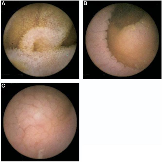
Background & aims: Wireless capsule endoscopy provides an opportunity to study the macroscopic features in celiac disease by providing a magnified view of the intestinal mucosa. In this study, we evaluated the following: (1) the distribution of atrophy in untreated celiac disease, (2) the correlation between extent of changes and clinical manifestations, (3) the accuracy and interobserver agreement of wireless capsule endoscopy assessment, and (4) the effect of gluten withdrawal.
Methods: Thirty-eight consecutive patients with untreated biopsy-proven celiac disease underwent wireless capsule endoscopy. Each subject was invited to undergo repeat testing after at least 6 months of gluten withdrawal. The video images of each patient were reviewed independently by 2 investigators.
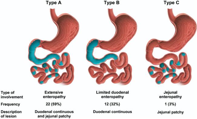
Results: Thirty-five (92%) subjects had visible atrophy detected by capsule endoscopy. Twenty-two (59%) subjects showed an extensive enteropathy, 12 (32%) had enteropathy limited to the duodenum, and only 1 had a jejunal enteropathy. No association was shown between the extent of the lesion and clinical manifestations. Capsule endoscopy had a better overall sensitivity for the detection of atrophy as compared with upper endoscopy (92% vs 55%, P = .0005), with a specificity of 100%. The overall interobserver agreement for the 2 reviewers was relatively high (% total agreement, 86.5%). After gluten withdrawal, the extent and the pattern of atrophy improved both qualitatively and quantitatively.
Conclusions: Celiac disease affects a highly variable portion of the small intestine starting at the duodenum. The extent of visible enteropathy does not explain differences in clinical presentation. Most subjects with visually detected villous atrophy showed a clinically significant improvement after gluten withdrawal.
Figures
Comment in
-
Mucosal atrophy extent and clinical correlation in celiac disease.Clin Gastroenterol Hepatol. 2008 Sep;6(9):1061; author reply 1061. doi: 10.1016/j.cgh.2008.02.057. Clin Gastroenterol Hepatol. 2008. PMID: 18774534 No abstract available.
References
-
- Rostom A, Murray JA, Kagnoff MF. American Gastroenterological Association (AGA) Institute technical review on diagnosis and management of celiac disease. Gastroenterology. 2006;131:1981–2002. - PubMed
-
- Ravelli AM, Tobanelli P, Minelli L, et al. Endoscopic features of celiac disease in children. Gastrointest Endosc. 2001;54:736–742. - PubMed
-
- Brar P, Kwon GY, Egbuna, et al. Lack of correlation of degree of villous atrophy with severity of clinical presentation of celiac disease. Dig Liver Dis. 2007;39:26–29. - PubMed
-
- Marsh MN, Crowe PT. Morphology of the mucosal lesion in gluten sensitivity. Baillieres Clin Gastroenterol. 1995;9:273–293. - PubMed
-
- MacDonald WC, Brandborg LL, Flich AL, et al. Studies of celiac sprue IV: the response of the whole length of the small bowel to a gluten free diet. Gastroenterology. 1964;47:573–589. - PubMed
Publication types
MeSH terms
Grants and funding
LinkOut - more resources
Full Text Sources
Medical
